Austin Crispin-Smith, Ti Wu, Ilana R Leppert, Agah Karakuzu, Shantanu Sinha, Usha Sinha
{"title":"评估磁化转移对比序列:应用于监测肌肉大分子分数的年龄相关差异。","authors":"Austin Crispin-Smith, Ti Wu, Ilana R Leppert, Agah Karakuzu, Shantanu Sinha, Usha Sinha","doi":"10.3390/tomography11090103","DOIUrl":null,"url":null,"abstract":"<p><strong>Background/objectives: </strong>Several sequences for magnetization transfer contrast (MTC) imaging are available, from indices of MTC ranging from quantitative magnetization transfer (qMT) that yields the macromolecular fraction to simple ratios of signal intensities with and without a magnetization transfer (MT) pulse. Aging muscle undergoes changes including an increase in fibrosis and adipose accompanied by fiber atrophy and loss. The objective is to evaluate five MTC sequences to study age-related differences in muscle tissue composition.</p><p><strong>Methods: </strong>The lower leg (calf) of 15 young (8M/7F, 25.8 ± 3.7 years) and 9 senior subjects (5F/4M, 68.4 ± 3.3 years) was imaged with the following sequences: multi-offset qMT fit to the Ramani and Yarnykh models, single-offset qMT two-parameter fit to the Ramani model, a semi-quantitative <i>MT<sub>sat</sub></i> sequence, magnetization transfer ratio (<i>MTR</i>), and MTR-corrected (<i>MTR<sub>corr</sub></i>) for B1 inhomogeneities. <i>T1</i> mapping was also performed. Statistical analysis was performed to identify significant age-related and regional (intermuscular) differences.</p><p><strong>Results: </strong>Significant age-related decreases (<i>p</i> < 0.001) in macromolecular fraction (from two-parameter fit), <i>MT<sub>sat</sub></i>, <i>MTR</i>, and <i>MTR<sub>corr</sub></i> were identified. A significant age-related increase in <i>T1</i> (<i>p</i> < 0.001) was also identified. Pearson correlation coefficients between <i>T1</i> and MTC indices were weak to moderate but significant.</p><p><strong>Conclusions: </strong>Age-related decreases in MTC may reflect that loss of myofibrillar proteins dominates the increase in collagen content with age. Further, the modest correlation of MTC indices with <i>T1</i> indicates that all the age-related differences in MTC cannot be explained by an increase in inflammation. The <i>MT<sub>sat</sub></i> sequence was identified as the most clinically relevant in terms of acquisition speed, post-processing simplicity, and ability to identify age-related differences in macromolecular fractions.</p>","PeriodicalId":51330,"journal":{"name":"Tomography","volume":"11 9","pages":""},"PeriodicalIF":2.2000,"publicationDate":"2025-09-05","publicationTypes":"Journal Article","fieldsOfStudy":null,"isOpenAccess":false,"openAccessPdf":"https://www.ncbi.nlm.nih.gov/pmc/articles/PMC12473340/pdf/","citationCount":"0","resultStr":"{\"title\":\"Evaluation of Magnetization Transfer Contrast Sequences: Application to Monitor Age-Related Differences in Muscle Macromolecular Fraction.\",\"authors\":\"Austin Crispin-Smith, Ti Wu, Ilana R Leppert, Agah Karakuzu, Shantanu Sinha, Usha Sinha\",\"doi\":\"10.3390/tomography11090103\",\"DOIUrl\":null,\"url\":null,\"abstract\":\"<p><strong>Background/objectives: </strong>Several sequences for magnetization transfer contrast (MTC) imaging are available, from indices of MTC ranging from quantitative magnetization transfer (qMT) that yields the macromolecular fraction to simple ratios of signal intensities with and without a magnetization transfer (MT) pulse. Aging muscle undergoes changes including an increase in fibrosis and adipose accompanied by fiber atrophy and loss. The objective is to evaluate five MTC sequences to study age-related differences in muscle tissue composition.</p><p><strong>Methods: </strong>The lower leg (calf) of 15 young (8M/7F, 25.8 ± 3.7 years) and 9 senior subjects (5F/4M, 68.4 ± 3.3 years) was imaged with the following sequences: multi-offset qMT fit to the Ramani and Yarnykh models, single-offset qMT two-parameter fit to the Ramani model, a semi-quantitative <i>MT<sub>sat</sub></i> sequence, magnetization transfer ratio (<i>MTR</i>), and MTR-corrected (<i>MTR<sub>corr</sub></i>) for B1 inhomogeneities. <i>T1</i> mapping was also performed. Statistical analysis was performed to identify significant age-related and regional (intermuscular) differences.</p><p><strong>Results: </strong>Significant age-related decreases (<i>p</i> < 0.001) in macromolecular fraction (from two-parameter fit), <i>MT<sub>sat</sub></i>, <i>MTR</i>, and <i>MTR<sub>corr</sub></i> were identified. A significant age-related increase in <i>T1</i> (<i>p</i> < 0.001) was also identified. Pearson correlation coefficients between <i>T1</i> and MTC indices were weak to moderate but significant.</p><p><strong>Conclusions: </strong>Age-related decreases in MTC may reflect that loss of myofibrillar proteins dominates the increase in collagen content with age. Further, the modest correlation of MTC indices with <i>T1</i> indicates that all the age-related differences in MTC cannot be explained by an increase in inflammation. The <i>MT<sub>sat</sub></i> sequence was identified as the most clinically relevant in terms of acquisition speed, post-processing simplicity, and ability to identify age-related differences in macromolecular fractions.</p>\",\"PeriodicalId\":51330,\"journal\":{\"name\":\"Tomography\",\"volume\":\"11 9\",\"pages\":\"\"},\"PeriodicalIF\":2.2000,\"publicationDate\":\"2025-09-05\",\"publicationTypes\":\"Journal Article\",\"fieldsOfStudy\":null,\"isOpenAccess\":false,\"openAccessPdf\":\"https://www.ncbi.nlm.nih.gov/pmc/articles/PMC12473340/pdf/\",\"citationCount\":\"0\",\"resultStr\":null,\"platform\":\"Semanticscholar\",\"paperid\":null,\"PeriodicalName\":\"Tomography\",\"FirstCategoryId\":\"3\",\"ListUrlMain\":\"https://doi.org/10.3390/tomography11090103\",\"RegionNum\":4,\"RegionCategory\":\"医学\",\"ArticlePicture\":[],\"TitleCN\":null,\"AbstractTextCN\":null,\"PMCID\":null,\"EPubDate\":\"\",\"PubModel\":\"\",\"JCR\":\"Q2\",\"JCRName\":\"RADIOLOGY, NUCLEAR MEDICINE & MEDICAL IMAGING\",\"Score\":null,\"Total\":0}","platform":"Semanticscholar","paperid":null,"PeriodicalName":"Tomography","FirstCategoryId":"3","ListUrlMain":"https://doi.org/10.3390/tomography11090103","RegionNum":4,"RegionCategory":"医学","ArticlePicture":[],"TitleCN":null,"AbstractTextCN":null,"PMCID":null,"EPubDate":"","PubModel":"","JCR":"Q2","JCRName":"RADIOLOGY, NUCLEAR MEDICINE & MEDICAL IMAGING","Score":null,"Total":0}
Evaluation of Magnetization Transfer Contrast Sequences: Application to Monitor Age-Related Differences in Muscle Macromolecular Fraction.
Background/objectives: Several sequences for magnetization transfer contrast (MTC) imaging are available, from indices of MTC ranging from quantitative magnetization transfer (qMT) that yields the macromolecular fraction to simple ratios of signal intensities with and without a magnetization transfer (MT) pulse. Aging muscle undergoes changes including an increase in fibrosis and adipose accompanied by fiber atrophy and loss. The objective is to evaluate five MTC sequences to study age-related differences in muscle tissue composition.
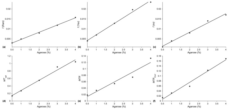
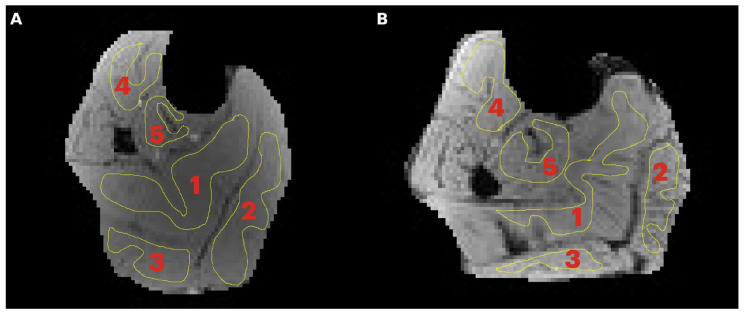
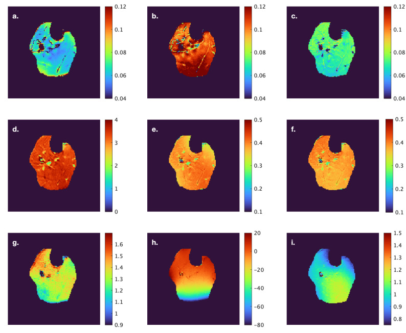
Methods: The lower leg (calf) of 15 young (8M/7F, 25.8 ± 3.7 years) and 9 senior subjects (5F/4M, 68.4 ± 3.3 years) was imaged with the following sequences: multi-offset qMT fit to the Ramani and Yarnykh models, single-offset qMT two-parameter fit to the Ramani model, a semi-quantitative MTsat sequence, magnetization transfer ratio (MTR), and MTR-corrected (MTRcorr) for B1 inhomogeneities. T1 mapping was also performed. Statistical analysis was performed to identify significant age-related and regional (intermuscular) differences.
Results: Significant age-related decreases (p < 0.001) in macromolecular fraction (from two-parameter fit), MTsat, MTR, and MTRcorr were identified. A significant age-related increase in T1 (p < 0.001) was also identified. Pearson correlation coefficients between T1 and MTC indices were weak to moderate but significant.
Conclusions: Age-related decreases in MTC may reflect that loss of myofibrillar proteins dominates the increase in collagen content with age. Further, the modest correlation of MTC indices with T1 indicates that all the age-related differences in MTC cannot be explained by an increase in inflammation. The MTsat sequence was identified as the most clinically relevant in terms of acquisition speed, post-processing simplicity, and ability to identify age-related differences in macromolecular fractions.
TomographyMedicine-Radiology, Nuclear Medicine and Imaging
CiteScore
2.70
自引率
10.50%
发文量
222
期刊介绍:
TomographyTM publishes basic (technical and pre-clinical) and clinical scientific articles which involve the advancement of imaging technologies. Tomography encompasses studies that use single or multiple imaging modalities including for example CT, US, PET, SPECT, MR and hyperpolarization technologies, as well as optical modalities (i.e. bioluminescence, photoacoustic, endomicroscopy, fiber optic imaging and optical computed tomography) in basic sciences, engineering, preclinical and clinical medicine.
Tomography also welcomes studies involving exploration and refinement of contrast mechanisms and image-derived metrics within and across modalities toward the development of novel imaging probes for image-based feedback and intervention. The use of imaging in biology and medicine provides unparalleled opportunities to noninvasively interrogate tissues to obtain real-time dynamic and quantitative information required for diagnosis and response to interventions and to follow evolving pathological conditions. As multi-modal studies and the complexities of imaging technologies themselves are ever increasing to provide advanced information to scientists and clinicians.
Tomography provides a unique publication venue allowing investigators the opportunity to more precisely communicate integrated findings related to the diverse and heterogeneous features associated with underlying anatomical, physiological, functional, metabolic and molecular genetic activities of normal and diseased tissue. Thus Tomography publishes peer-reviewed articles which involve the broad use of imaging of any tissue and disease type including both preclinical and clinical investigations. In addition, hardware/software along with chemical and molecular probe advances are welcome as they are deemed to significantly contribute towards the long-term goal of improving the overall impact of imaging on scientific and clinical discovery.

 求助内容:
求助内容: 应助结果提醒方式:


