Leila Pereira Tenório, José Batista Volpon, Marcello Henrique Nogueira-Barbosa
{"title":"小儿软骨髌骨的超声检查:一个病例系列。","authors":"Leila Pereira Tenório, José Batista Volpon, Marcello Henrique Nogueira-Barbosa","doi":"10.1590/1413-785220253305e291176","DOIUrl":null,"url":null,"abstract":"<p><strong>Objective: </strong>To analyze cases with clinical suspicion of patellar abnormalities, before ossification of the patella and to characterize the spectrum of abnormalities of the cartilaginous infantile patella by ultrasonography.</p><p><strong>Methods: </strong>Retrospective study using the keyword \"patella\" in ultrasonography reports in the Radiology Information System (RIS). The main researcher performed patellar measurements in the group of patients and in a control group (9 patients) without clinical or ultrasonography abnormalities.</p><p><strong>Results: </strong>Twelve patients with suspected patellar abnormalities were identified, with a mean age of 9 months and 4 days (±1.9 years), 75% male.</p><p><strong>Findings: </strong>dislocation or subluxation associated with patellar hypoplasia (7 knees), low lying patella and patellar hypoplasia (2), unilateral patellar agenesis (1), bilateral patellar agenesis (1), patellar instability in dynamic assessment and absence of patellar morphological changes (1). In two patients, ultrasonography was negative. The craniocaudal diameter of the hypoplastic patellas measured 0.94 cm ± 0.24 cm and in the control group 1.24 cm and ±0.12 cm (p<0.01). The Insall-Salvati index adapted for ultrasonography measured 0.63±0.07 for the low lying patella and 0.93±0.16 in the control group (p=0.004).</p><p><strong>Conclusions: </strong>Ultrasonography was useful to characterize abnormalities of the cartilaginous patella, and the most frequent findings were instability and hypoplasia. <b><i>Level of Evidence IV; Case Series.</i></b></p>","PeriodicalId":55563,"journal":{"name":"Acta Ortopedica Brasileira","volume":"33 5","pages":"e291176"},"PeriodicalIF":0.6000,"publicationDate":"2025-09-22","publicationTypes":"Journal Article","fieldsOfStudy":null,"isOpenAccess":false,"openAccessPdf":"https://www.ncbi.nlm.nih.gov/pmc/articles/PMC12456895/pdf/","citationCount":"0","resultStr":"{\"title\":\"ULTRASONOGRAPHY OF THE CARTILAGINOUS PATELLA IN PEDIATRIC PATIENTS: A CASE SERIES.\",\"authors\":\"Leila Pereira Tenório, José Batista Volpon, Marcello Henrique Nogueira-Barbosa\",\"doi\":\"10.1590/1413-785220253305e291176\",\"DOIUrl\":null,\"url\":null,\"abstract\":\"<p><strong>Objective: </strong>To analyze cases with clinical suspicion of patellar abnormalities, before ossification of the patella and to characterize the spectrum of abnormalities of the cartilaginous infantile patella by ultrasonography.</p><p><strong>Methods: </strong>Retrospective study using the keyword \\\"patella\\\" in ultrasonography reports in the Radiology Information System (RIS). The main researcher performed patellar measurements in the group of patients and in a control group (9 patients) without clinical or ultrasonography abnormalities.</p><p><strong>Results: </strong>Twelve patients with suspected patellar abnormalities were identified, with a mean age of 9 months and 4 days (±1.9 years), 75% male.</p><p><strong>Findings: </strong>dislocation or subluxation associated with patellar hypoplasia (7 knees), low lying patella and patellar hypoplasia (2), unilateral patellar agenesis (1), bilateral patellar agenesis (1), patellar instability in dynamic assessment and absence of patellar morphological changes (1). In two patients, ultrasonography was negative. The craniocaudal diameter of the hypoplastic patellas measured 0.94 cm ± 0.24 cm and in the control group 1.24 cm and ±0.12 cm (p<0.01). The Insall-Salvati index adapted for ultrasonography measured 0.63±0.07 for the low lying patella and 0.93±0.16 in the control group (p=0.004).</p><p><strong>Conclusions: </strong>Ultrasonography was useful to characterize abnormalities of the cartilaginous patella, and the most frequent findings were instability and hypoplasia. <b><i>Level of Evidence IV; Case Series.</i></b></p>\",\"PeriodicalId\":55563,\"journal\":{\"name\":\"Acta Ortopedica Brasileira\",\"volume\":\"33 5\",\"pages\":\"e291176\"},\"PeriodicalIF\":0.6000,\"publicationDate\":\"2025-09-22\",\"publicationTypes\":\"Journal Article\",\"fieldsOfStudy\":null,\"isOpenAccess\":false,\"openAccessPdf\":\"https://www.ncbi.nlm.nih.gov/pmc/articles/PMC12456895/pdf/\",\"citationCount\":\"0\",\"resultStr\":null,\"platform\":\"Semanticscholar\",\"paperid\":null,\"PeriodicalName\":\"Acta Ortopedica Brasileira\",\"FirstCategoryId\":\"3\",\"ListUrlMain\":\"https://doi.org/10.1590/1413-785220253305e291176\",\"RegionNum\":4,\"RegionCategory\":\"医学\",\"ArticlePicture\":[],\"TitleCN\":null,\"AbstractTextCN\":null,\"PMCID\":null,\"EPubDate\":\"2025/1/1 0:00:00\",\"PubModel\":\"eCollection\",\"JCR\":\"Q4\",\"JCRName\":\"ORTHOPEDICS\",\"Score\":null,\"Total\":0}","platform":"Semanticscholar","paperid":null,"PeriodicalName":"Acta Ortopedica Brasileira","FirstCategoryId":"3","ListUrlMain":"https://doi.org/10.1590/1413-785220253305e291176","RegionNum":4,"RegionCategory":"医学","ArticlePicture":[],"TitleCN":null,"AbstractTextCN":null,"PMCID":null,"EPubDate":"2025/1/1 0:00:00","PubModel":"eCollection","JCR":"Q4","JCRName":"ORTHOPEDICS","Score":null,"Total":0}
ULTRASONOGRAPHY OF THE CARTILAGINOUS PATELLA IN PEDIATRIC PATIENTS: A CASE SERIES.
Objective: To analyze cases with clinical suspicion of patellar abnormalities, before ossification of the patella and to characterize the spectrum of abnormalities of the cartilaginous infantile patella by ultrasonography.
Methods: Retrospective study using the keyword "patella" in ultrasonography reports in the Radiology Information System (RIS). The main researcher performed patellar measurements in the group of patients and in a control group (9 patients) without clinical or ultrasonography abnormalities.
Results: Twelve patients with suspected patellar abnormalities were identified, with a mean age of 9 months and 4 days (±1.9 years), 75% male.
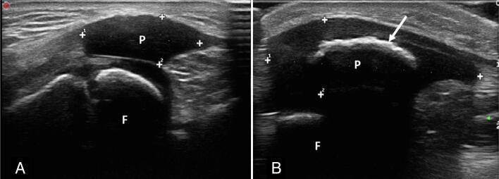
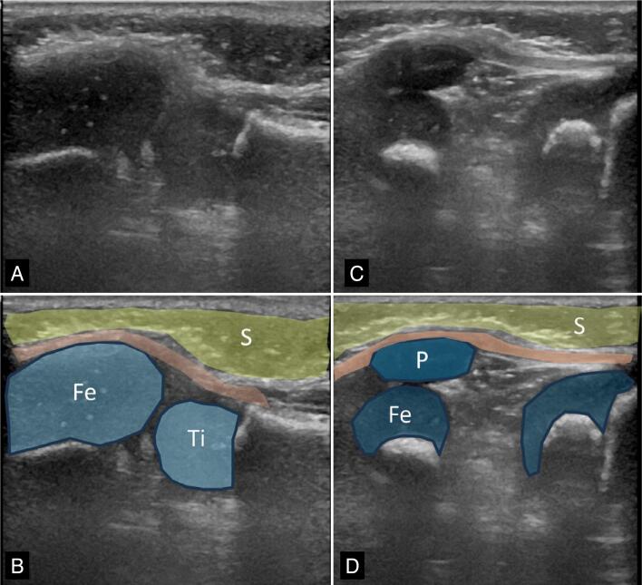
Findings: dislocation or subluxation associated with patellar hypoplasia (7 knees), low lying patella and patellar hypoplasia (2), unilateral patellar agenesis (1), bilateral patellar agenesis (1), patellar instability in dynamic assessment and absence of patellar morphological changes (1). In two patients, ultrasonography was negative. The craniocaudal diameter of the hypoplastic patellas measured 0.94 cm ± 0.24 cm and in the control group 1.24 cm and ±0.12 cm (p<0.01). The Insall-Salvati index adapted for ultrasonography measured 0.63±0.07 for the low lying patella and 0.93±0.16 in the control group (p=0.004).
Conclusions: Ultrasonography was useful to characterize abnormalities of the cartilaginous patella, and the most frequent findings were instability and hypoplasia. Level of Evidence IV; Case Series.
期刊介绍:
A Revista Acta Ortopédica Brasileira, órgão oficial do Departamento de Ortopedia e Traumatologia da Faculdade de Medicina da Universidade de São Paulo (DOT/FMUSP), é publicada bimestralmente em seis edições ao ano (jan/fev, mar/abr, maio/jun, jul/ago, set/out e nov/dez) com versão em inglês disponível nos principais indexadores nacionais e internacionais e instituições de ensino do Brasil. Sendo hoje reconhecidamente uma importante contribuição para os especialistas da área com sua seriedade e árduo trabalho para as indexações já conquistadas.


 求助内容:
求助内容: 应助结果提醒方式:


