Frederik Fuchs, Sebastian N Marschner, Jan Hofmaier, Maya Rottler, Indra Hadi, Sebastian H Maier, Tobias Greve, Adrien Holzgreve, Nathalie L Albert, Raphael Bodensohn, Claus Belka, Maximilian Niyazi, Franziska Walter
{"title":"颅底低级别脑膜瘤的SSTR PET/CT:放射治疗中精确大体肿瘤体积描绘的关键工具?","authors":"Frederik Fuchs, Sebastian N Marschner, Jan Hofmaier, Maya Rottler, Indra Hadi, Sebastian H Maier, Tobias Greve, Adrien Holzgreve, Nathalie L Albert, Raphael Bodensohn, Claus Belka, Maximilian Niyazi, Franziska Walter","doi":"10.1186/s13014-025-02718-4","DOIUrl":null,"url":null,"abstract":"<p><strong>Background: </strong>Precise delineation of gross tumor volume (GTV) is fundamental for effective radiation therapy in low-grade skull base meningiomas. Magnetic resonance imaging (MRI) serves as the primary imaging tool but may not fully represent tumor extent. This study investigates the additional value of incorporating Somatostatin receptor (SSTR)-directed PET/CT in radiation therapy planning.</p><p><strong>Methods: </strong>A retrospective analysis was conducted with four experienced radiation oncologists contouring GTVs for skull base meningiomas using MRI alone (GTV_MRI), PET/CT alone (GTV_PET/CT), and both modalities combined (GTV_ALL). Consensus ground truth volumes were generated for each modality through a STAPLE algorithm. Agreement between modalities, excluding observer variability, was assessed using statistical metrics including Dice Similarity Coefficient (DSC), Jaccard Index (JCI), Hausdorff distance (HD95), Geographical Miss Index (GMI), sensitivity, and kappa statistics.</p><p><strong>Results: </strong>The study included 25 patients (15 females, 10 males; median age 56 years (range: 23-74 years), with 96% achieving local control post-radiotherapy over a median follow-up of 64 months (range: 28-135 months). Substantial interobserver agreement was observed, with median kappa values of 0.74 for GTV_MRI, 0.68 for GTV_PET/CT, and 0.77 for GTV_ALL. Median consensus volumes were 6.65 cc (MRI<sub>STAPLE</sub>), 7.21 cc (PET<sub>STAPLE</sub>), and 6.73 cc (ALL<sub>STAPLE</sub>). The median GMI for MRI<sub>STAPLE</sub> compared to ALL<sub>STAPLE</sub> was 0.18 (IQR: 0.11-0.39), and 0.21 (IQR: 0.15-0.28) for PET<sub>STAPLE</sub> compared to ALL<sub>STAPLE</sub>. The DSC indicated the lowest concordance between MRI<sub>STAPLE</sub> and PET<sub>STAPLE</sub> with a median of 0.75 (IQR: 0.59-0.82), followed by PET<sub>STAPLE</sub> versus ALL<sub>STAPLE</sub> with a median DSC of 0.84 (IQR: 0.79-0.89), and MRI<sub>STAPLE</sub> versus ALL<sub>STAPLE</sub> with a median DSC of 0.89 (IQR: 0.76-0.92). The integration of PET/CT with MRI significantly enhanced concordance metrics.</p><p><strong>Conclusion: </strong>Combining MRI and PET/CT improves GTV delineation in low-grade skull base meningiomas, as PET/CT can reveal regions missed by MRI, which may slightly underestimate tumor size. This multimodal imaging approach enhances consensus and supports its role in radiotherapy planning. Standardized protocols and technical integration remain key future goals.</p>","PeriodicalId":49639,"journal":{"name":"Radiation Oncology","volume":"20 1","pages":"142"},"PeriodicalIF":3.3000,"publicationDate":"2025-09-22","publicationTypes":"Journal Article","fieldsOfStudy":null,"isOpenAccess":false,"openAccessPdf":"https://www.ncbi.nlm.nih.gov/pmc/articles/PMC12455788/pdf/","citationCount":"0","resultStr":"{\"title\":\"SSTR PET/CT for skull base low-grade meningioma: a critical tool for accurate gross tumor volume delineation in radiotherapy?\",\"authors\":\"Frederik Fuchs, Sebastian N Marschner, Jan Hofmaier, Maya Rottler, Indra Hadi, Sebastian H Maier, Tobias Greve, Adrien Holzgreve, Nathalie L Albert, Raphael Bodensohn, Claus Belka, Maximilian Niyazi, Franziska Walter\",\"doi\":\"10.1186/s13014-025-02718-4\",\"DOIUrl\":null,\"url\":null,\"abstract\":\"<p><strong>Background: </strong>Precise delineation of gross tumor volume (GTV) is fundamental for effective radiation therapy in low-grade skull base meningiomas. Magnetic resonance imaging (MRI) serves as the primary imaging tool but may not fully represent tumor extent. This study investigates the additional value of incorporating Somatostatin receptor (SSTR)-directed PET/CT in radiation therapy planning.</p><p><strong>Methods: </strong>A retrospective analysis was conducted with four experienced radiation oncologists contouring GTVs for skull base meningiomas using MRI alone (GTV_MRI), PET/CT alone (GTV_PET/CT), and both modalities combined (GTV_ALL). Consensus ground truth volumes were generated for each modality through a STAPLE algorithm. Agreement between modalities, excluding observer variability, was assessed using statistical metrics including Dice Similarity Coefficient (DSC), Jaccard Index (JCI), Hausdorff distance (HD95), Geographical Miss Index (GMI), sensitivity, and kappa statistics.</p><p><strong>Results: </strong>The study included 25 patients (15 females, 10 males; median age 56 years (range: 23-74 years), with 96% achieving local control post-radiotherapy over a median follow-up of 64 months (range: 28-135 months). Substantial interobserver agreement was observed, with median kappa values of 0.74 for GTV_MRI, 0.68 for GTV_PET/CT, and 0.77 for GTV_ALL. Median consensus volumes were 6.65 cc (MRI<sub>STAPLE</sub>), 7.21 cc (PET<sub>STAPLE</sub>), and 6.73 cc (ALL<sub>STAPLE</sub>). The median GMI for MRI<sub>STAPLE</sub> compared to ALL<sub>STAPLE</sub> was 0.18 (IQR: 0.11-0.39), and 0.21 (IQR: 0.15-0.28) for PET<sub>STAPLE</sub> compared to ALL<sub>STAPLE</sub>. The DSC indicated the lowest concordance between MRI<sub>STAPLE</sub> and PET<sub>STAPLE</sub> with a median of 0.75 (IQR: 0.59-0.82), followed by PET<sub>STAPLE</sub> versus ALL<sub>STAPLE</sub> with a median DSC of 0.84 (IQR: 0.79-0.89), and MRI<sub>STAPLE</sub> versus ALL<sub>STAPLE</sub> with a median DSC of 0.89 (IQR: 0.76-0.92). The integration of PET/CT with MRI significantly enhanced concordance metrics.</p><p><strong>Conclusion: </strong>Combining MRI and PET/CT improves GTV delineation in low-grade skull base meningiomas, as PET/CT can reveal regions missed by MRI, which may slightly underestimate tumor size. This multimodal imaging approach enhances consensus and supports its role in radiotherapy planning. Standardized protocols and technical integration remain key future goals.</p>\",\"PeriodicalId\":49639,\"journal\":{\"name\":\"Radiation Oncology\",\"volume\":\"20 1\",\"pages\":\"142\"},\"PeriodicalIF\":3.3000,\"publicationDate\":\"2025-09-22\",\"publicationTypes\":\"Journal Article\",\"fieldsOfStudy\":null,\"isOpenAccess\":false,\"openAccessPdf\":\"https://www.ncbi.nlm.nih.gov/pmc/articles/PMC12455788/pdf/\",\"citationCount\":\"0\",\"resultStr\":null,\"platform\":\"Semanticscholar\",\"paperid\":null,\"PeriodicalName\":\"Radiation Oncology\",\"FirstCategoryId\":\"3\",\"ListUrlMain\":\"https://doi.org/10.1186/s13014-025-02718-4\",\"RegionNum\":2,\"RegionCategory\":\"医学\",\"ArticlePicture\":[],\"TitleCN\":null,\"AbstractTextCN\":null,\"PMCID\":null,\"EPubDate\":\"\",\"PubModel\":\"\",\"JCR\":\"Q2\",\"JCRName\":\"ONCOLOGY\",\"Score\":null,\"Total\":0}","platform":"Semanticscholar","paperid":null,"PeriodicalName":"Radiation Oncology","FirstCategoryId":"3","ListUrlMain":"https://doi.org/10.1186/s13014-025-02718-4","RegionNum":2,"RegionCategory":"医学","ArticlePicture":[],"TitleCN":null,"AbstractTextCN":null,"PMCID":null,"EPubDate":"","PubModel":"","JCR":"Q2","JCRName":"ONCOLOGY","Score":null,"Total":0}
引用次数: 0
摘要
背景:准确描绘总肿瘤体积(GTV)是低级别颅底脑膜瘤有效放射治疗的基础。磁共振成像(MRI)是主要的成像工具,但可能不能完全代表肿瘤的范围。本研究探讨了结合生长抑素受体(SSTR)定向PET/CT在放射治疗计划中的附加价值。方法:回顾性分析4名经验丰富的放射肿瘤学家分别使用MRI (GTV_MRI)、PET/CT (GTV_PET/CT)和两种方式联合(GTV_ALL)对颅底脑膜瘤进行gtv轮廓的临床资料。通过STAPLE算法为每种模态生成共识基础真量。采用统计指标,包括Dice Similarity Coefficient (DSC)、Jaccard Index (JCI)、Hausdorff distance (HD95)、Geographical Miss Index (GMI)、sensitivity(灵敏度)和kappa statistics,评估模式之间的一致性,排除观察者的可变性。结果:本研究纳入25例患者,其中女性15例,男性10例,中位年龄56岁(范围23-74岁),96%患者放疗后局部控制,中位随访64个月(范围28-135个月)。观察到大量观察者之间的一致,GTV_MRI的中位kappa值为0.74,GTV_PET/CT为0.68,GTV_ALL为0.77。中位共识容积为6.65 cc (mrristaple)、7.21 cc (PETSTAPLE)和6.73 cc (ALLSTAPLE)。与ALLSTAPLE相比,mri的中位GMI为0.18 (IQR: 0.11-0.39), PETSTAPLE与ALLSTAPLE的中位GMI为0.21 (IQR: 0.15-0.28)。DSC显示,mri与PETSTAPLE的一致性最低,中位数为0.75 (IQR: 0.59-0.82),其次是PETSTAPLE与ALLSTAPLE的中位数DSC为0.84 (IQR: 0.79-0.89), mri与ALLSTAPLE的中位数DSC为0.89 (IQR: 0.76-0.92)。PET/CT与MRI的整合显著增强了一致性指标。结论:MRI与PET/CT结合可以改善低级别颅底脑膜瘤的GTV描绘,因为PET/CT可以显示MRI遗漏的区域,可能略低估肿瘤大小。这种多模态成像方法增强了共识,并支持其在放疗计划中的作用。标准化协议和技术集成仍然是未来的关键目标。
SSTR PET/CT for skull base low-grade meningioma: a critical tool for accurate gross tumor volume delineation in radiotherapy?
Background: Precise delineation of gross tumor volume (GTV) is fundamental for effective radiation therapy in low-grade skull base meningiomas. Magnetic resonance imaging (MRI) serves as the primary imaging tool but may not fully represent tumor extent. This study investigates the additional value of incorporating Somatostatin receptor (SSTR)-directed PET/CT in radiation therapy planning.
Methods: A retrospective analysis was conducted with four experienced radiation oncologists contouring GTVs for skull base meningiomas using MRI alone (GTV_MRI), PET/CT alone (GTV_PET/CT), and both modalities combined (GTV_ALL). Consensus ground truth volumes were generated for each modality through a STAPLE algorithm. Agreement between modalities, excluding observer variability, was assessed using statistical metrics including Dice Similarity Coefficient (DSC), Jaccard Index (JCI), Hausdorff distance (HD95), Geographical Miss Index (GMI), sensitivity, and kappa statistics.
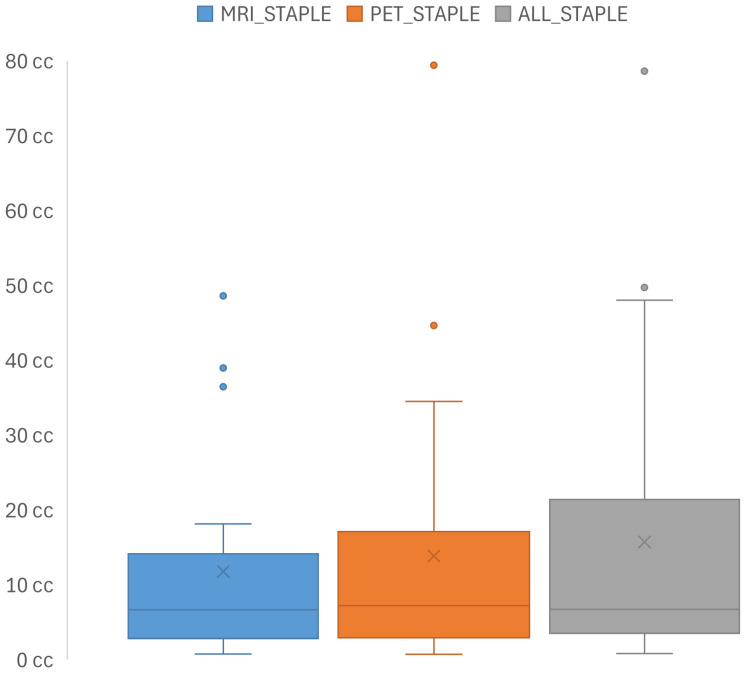
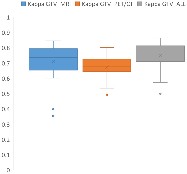
Results: The study included 25 patients (15 females, 10 males; median age 56 years (range: 23-74 years), with 96% achieving local control post-radiotherapy over a median follow-up of 64 months (range: 28-135 months). Substantial interobserver agreement was observed, with median kappa values of 0.74 for GTV_MRI, 0.68 for GTV_PET/CT, and 0.77 for GTV_ALL. Median consensus volumes were 6.65 cc (MRISTAPLE), 7.21 cc (PETSTAPLE), and 6.73 cc (ALLSTAPLE). The median GMI for MRISTAPLE compared to ALLSTAPLE was 0.18 (IQR: 0.11-0.39), and 0.21 (IQR: 0.15-0.28) for PETSTAPLE compared to ALLSTAPLE. The DSC indicated the lowest concordance between MRISTAPLE and PETSTAPLE with a median of 0.75 (IQR: 0.59-0.82), followed by PETSTAPLE versus ALLSTAPLE with a median DSC of 0.84 (IQR: 0.79-0.89), and MRISTAPLE versus ALLSTAPLE with a median DSC of 0.89 (IQR: 0.76-0.92). The integration of PET/CT with MRI significantly enhanced concordance metrics.
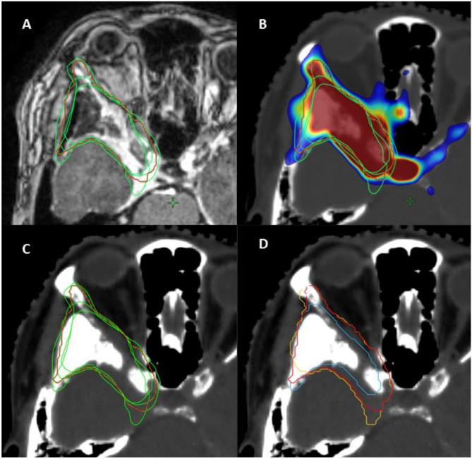
Conclusion: Combining MRI and PET/CT improves GTV delineation in low-grade skull base meningiomas, as PET/CT can reveal regions missed by MRI, which may slightly underestimate tumor size. This multimodal imaging approach enhances consensus and supports its role in radiotherapy planning. Standardized protocols and technical integration remain key future goals.
Radiation OncologyONCOLOGY-RADIOLOGY, NUCLEAR MEDICINE & MEDICAL IMAGING
CiteScore
6.50
自引率
2.80%
发文量
181
审稿时长
3-6 weeks
期刊介绍:
Radiation Oncology encompasses all aspects of research that impacts on the treatment of cancer using radiation. It publishes findings in molecular and cellular radiation biology, radiation physics, radiation technology, and clinical oncology.
 求助内容:
求助内容: 应助结果提醒方式:


