注意角落:在冷冻fib薄片制备过程中,将薄片因应力集中和薄片破裂造成的样品损失降到最低
IF 2.7
3区 生物学
Q3 BIOCHEMISTRY & MOLECULAR BIOLOGY
引用次数: 0
摘要
生物标本的冷冻fib铣削是冷冻电子断层成像工作流程中的关键和限制步骤。制备电子透明冷晶片是一个连续的、低通量的过程。即使采用自动化,熟练的操作人员通常也只能在单次冷冻fib会话中生产15-25片薄片。在样品处理、铣削和转移过程中,由于温度波动,冷冻固定细胞以及支撑膜层面临各种机械力和热应力。此外,细胞经过低温fib铣削后,产生的薄片继续承受机械处理和热应力的外力。我们提出了一个简单的,但非常有效的修改标准的矩形铣削模式通过实施“圆角”或角平滑提供更好的机械稳定性。这种调整有助于避免在薄片边缘的尖角,从而减少应力集中。结果,实验证实,这种改性降低了片层断裂的可能性,并将准备透射电镜的片层的总收率提高了40%以上。本文章由计算机程序翻译,如有差异,请以英文原文为准。
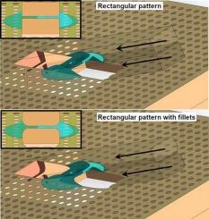
Mind the corner: Fillets in cryo-FIB lamella preparation to minimise sample loss caused by stress concentration and lamella breakage
Cryo-FIB milling of biological specimens is a critical and limiting step in the cryo-electron tomography workflow. Preparing electron-transparent cryo-lamellae is a serial, low-throughput process. Even with automation, a skilled operator can typically only produce 15–25 lamellae in a single cryo-FIB session. During sample handling, milling and transfer, the cryo-fixed cells as well as the supporting film layer face various mechanical forces and thermal stresses due to temperature fluctuations. Moreover, after cells are cryo-FIB milled, the resulting thin lamellae continue to endure external forces from mechanical handling and thermal stress. We propose a simple, yet highly effective modification to the standard rectangular milling pattern by implementing “fillets” or corner smoothing providing better mechanical stability. This adjustment helps to avoid sharp corners at the lamella edges, thereby reducing stress concentration. As a result, this modification decreases the likelihood of lamella breakage and improves the overall yield of ready-for-TEM lamellae by over 40 % as verified experimentally.
求助全文
通过发布文献求助,成功后即可免费获取论文全文。
去求助
来源期刊

Journal of structural biology
生物-生化与分子生物学
CiteScore
6.30
自引率
3.30%
发文量
88
审稿时长
65 days
期刊介绍:
Journal of Structural Biology (JSB) has an open access mirror journal, the Journal of Structural Biology: X (JSBX), sharing the same aims and scope, editorial team, submission system and rigorous peer review. Since both journals share the same editorial system, you may submit your manuscript via either journal homepage. You will be prompted during submission (and revision) to choose in which to publish your article. The editors and reviewers are not aware of the choice you made until the article has been published online. JSB and JSBX publish papers dealing with the structural analysis of living material at every level of organization by all methods that lead to an understanding of biological function in terms of molecular and supermolecular structure.
Techniques covered include:
• Light microscopy including confocal microscopy
• All types of electron microscopy
• X-ray diffraction
• Nuclear magnetic resonance
• Scanning force microscopy, scanning probe microscopy, and tunneling microscopy
• Digital image processing
• Computational insights into structure
 求助内容:
求助内容: 应助结果提醒方式:


