Paolo Bosco, Irina Podda, Emilio Cipriano, Clara Bombonato, Paola Cipriani, Mariaelisa Bartoli, Rosa Pasquariello, Simona Fiori, Laura Biagi, Anna Chilosi
{"title":"儿童言语失用症的神经基质改变:来自神经影像学的新证据。","authors":"Paolo Bosco, Irina Podda, Emilio Cipriano, Clara Bombonato, Paola Cipriani, Mariaelisa Bartoli, Rosa Pasquariello, Simona Fiori, Laura Biagi, Anna Chilosi","doi":"10.1093/braincomms/fcaf302","DOIUrl":null,"url":null,"abstract":"<p><p>Childhood apraxia of speech is a motor speech disorder characterized by deficits in programming, planning, and movement control, necessary for speech production with severe impact on oral and written language. Currently there are few studies on how speech is functionally rooted in neuroanatomy in children with apraxia of speech. The present cross-sectional study aimed at further identifying the specific neuroanatomical substrate of childhood apraxia of speech and at analysing the relationship between Magnetic Resonance Imaging findings and speech measures in a relatively large group of Italian children with this disorder. The brain structures of 71 children with apraxia of speech were compared to those of 30 age-matched typically developing peers. For each subject, the morphological brain images were segmented according to a specific atlas, and cortical thickness and volume measures were extracted from cortical and subcortical structures, respectively. Moreover, using voxel-based morphometry with a diffeomorphic anatomical registration procedure, grey matter morphometry of the two groups was compared. We also used diffusion weighted imaging (in 67 out of 71 children with apraxia of speech and all typically developing peers) to investigate the white matter integrity in specific speech-language-related tracts, comparing the mean fractional anisotropy along the tracts. Children with apraxia showed significantly greater grey matter volumes and increased cortical thickness than their typically developing peers in several areas involved in speech and language processing. After correction for multiple comparisons, significant increments in cortical thickness and volume survived in the left postcentral gyrus and bilaterally in the thalami. The diffusion study revealed a significant reduction of fractional anisotropy in childhood apraxia of speech with respect to typically developing children in the left frontal aslant tract in both supplementary motor area and pre-supplementary motor components. Speech severity and diadochokinetic rate of children with apraxia of speech correlated significantly with cortical thickness and volume measures extracted from the rostral middle-frontal gyrus, the left precuneus, and the left thalamus. The same speech measures correlated also with the fractional anisotropy values along the frontal aslant tract. Our results revealed grey matter alterations in childhood apraxia of speech, in a widespread circuit of cortical and subcortical areas, and in particular in both thalami and the left postcentral gyrus, crucial regions for sensorimotor control. Moreover, diffusion study results also provided further support to the involvement, in childhood apraxia of speech, of the left frontal aslant tract, a fundamental pathway for speech movement planning and programming.</p>","PeriodicalId":93915,"journal":{"name":"Brain communications","volume":"7 5","pages":"fcaf302"},"PeriodicalIF":4.5000,"publicationDate":"2025-09-16","publicationTypes":"Journal Article","fieldsOfStudy":null,"isOpenAccess":false,"openAccessPdf":"https://www.ncbi.nlm.nih.gov/pmc/articles/PMC12448706/pdf/","citationCount":"0","resultStr":"{\"title\":\"Alterations of the neural substrate in childhood apraxia of speech: new evidence from neuroimaging.\",\"authors\":\"Paolo Bosco, Irina Podda, Emilio Cipriano, Clara Bombonato, Paola Cipriani, Mariaelisa Bartoli, Rosa Pasquariello, Simona Fiori, Laura Biagi, Anna Chilosi\",\"doi\":\"10.1093/braincomms/fcaf302\",\"DOIUrl\":null,\"url\":null,\"abstract\":\"<p><p>Childhood apraxia of speech is a motor speech disorder characterized by deficits in programming, planning, and movement control, necessary for speech production with severe impact on oral and written language. Currently there are few studies on how speech is functionally rooted in neuroanatomy in children with apraxia of speech. The present cross-sectional study aimed at further identifying the specific neuroanatomical substrate of childhood apraxia of speech and at analysing the relationship between Magnetic Resonance Imaging findings and speech measures in a relatively large group of Italian children with this disorder. The brain structures of 71 children with apraxia of speech were compared to those of 30 age-matched typically developing peers. For each subject, the morphological brain images were segmented according to a specific atlas, and cortical thickness and volume measures were extracted from cortical and subcortical structures, respectively. Moreover, using voxel-based morphometry with a diffeomorphic anatomical registration procedure, grey matter morphometry of the two groups was compared. We also used diffusion weighted imaging (in 67 out of 71 children with apraxia of speech and all typically developing peers) to investigate the white matter integrity in specific speech-language-related tracts, comparing the mean fractional anisotropy along the tracts. Children with apraxia showed significantly greater grey matter volumes and increased cortical thickness than their typically developing peers in several areas involved in speech and language processing. After correction for multiple comparisons, significant increments in cortical thickness and volume survived in the left postcentral gyrus and bilaterally in the thalami. The diffusion study revealed a significant reduction of fractional anisotropy in childhood apraxia of speech with respect to typically developing children in the left frontal aslant tract in both supplementary motor area and pre-supplementary motor components. Speech severity and diadochokinetic rate of children with apraxia of speech correlated significantly with cortical thickness and volume measures extracted from the rostral middle-frontal gyrus, the left precuneus, and the left thalamus. The same speech measures correlated also with the fractional anisotropy values along the frontal aslant tract. Our results revealed grey matter alterations in childhood apraxia of speech, in a widespread circuit of cortical and subcortical areas, and in particular in both thalami and the left postcentral gyrus, crucial regions for sensorimotor control. Moreover, diffusion study results also provided further support to the involvement, in childhood apraxia of speech, of the left frontal aslant tract, a fundamental pathway for speech movement planning and programming.</p>\",\"PeriodicalId\":93915,\"journal\":{\"name\":\"Brain communications\",\"volume\":\"7 5\",\"pages\":\"fcaf302\"},\"PeriodicalIF\":4.5000,\"publicationDate\":\"2025-09-16\",\"publicationTypes\":\"Journal Article\",\"fieldsOfStudy\":null,\"isOpenAccess\":false,\"openAccessPdf\":\"https://www.ncbi.nlm.nih.gov/pmc/articles/PMC12448706/pdf/\",\"citationCount\":\"0\",\"resultStr\":null,\"platform\":\"Semanticscholar\",\"paperid\":null,\"PeriodicalName\":\"Brain communications\",\"FirstCategoryId\":\"1085\",\"ListUrlMain\":\"https://doi.org/10.1093/braincomms/fcaf302\",\"RegionNum\":0,\"RegionCategory\":null,\"ArticlePicture\":[],\"TitleCN\":null,\"AbstractTextCN\":null,\"PMCID\":null,\"EPubDate\":\"2025/1/1 0:00:00\",\"PubModel\":\"eCollection\",\"JCR\":\"Q1\",\"JCRName\":\"CLINICAL NEUROLOGY\",\"Score\":null,\"Total\":0}","platform":"Semanticscholar","paperid":null,"PeriodicalName":"Brain communications","FirstCategoryId":"1085","ListUrlMain":"https://doi.org/10.1093/braincomms/fcaf302","RegionNum":0,"RegionCategory":null,"ArticlePicture":[],"TitleCN":null,"AbstractTextCN":null,"PMCID":null,"EPubDate":"2025/1/1 0:00:00","PubModel":"eCollection","JCR":"Q1","JCRName":"CLINICAL NEUROLOGY","Score":null,"Total":0}
Alterations of the neural substrate in childhood apraxia of speech: new evidence from neuroimaging.
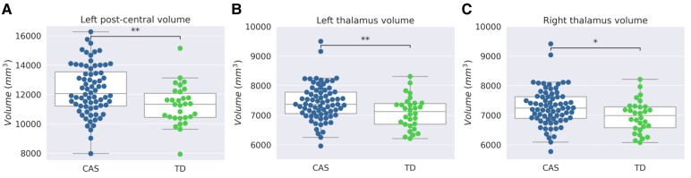
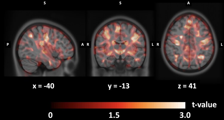
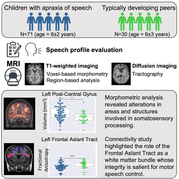
Childhood apraxia of speech is a motor speech disorder characterized by deficits in programming, planning, and movement control, necessary for speech production with severe impact on oral and written language. Currently there are few studies on how speech is functionally rooted in neuroanatomy in children with apraxia of speech. The present cross-sectional study aimed at further identifying the specific neuroanatomical substrate of childhood apraxia of speech and at analysing the relationship between Magnetic Resonance Imaging findings and speech measures in a relatively large group of Italian children with this disorder. The brain structures of 71 children with apraxia of speech were compared to those of 30 age-matched typically developing peers. For each subject, the morphological brain images were segmented according to a specific atlas, and cortical thickness and volume measures were extracted from cortical and subcortical structures, respectively. Moreover, using voxel-based morphometry with a diffeomorphic anatomical registration procedure, grey matter morphometry of the two groups was compared. We also used diffusion weighted imaging (in 67 out of 71 children with apraxia of speech and all typically developing peers) to investigate the white matter integrity in specific speech-language-related tracts, comparing the mean fractional anisotropy along the tracts. Children with apraxia showed significantly greater grey matter volumes and increased cortical thickness than their typically developing peers in several areas involved in speech and language processing. After correction for multiple comparisons, significant increments in cortical thickness and volume survived in the left postcentral gyrus and bilaterally in the thalami. The diffusion study revealed a significant reduction of fractional anisotropy in childhood apraxia of speech with respect to typically developing children in the left frontal aslant tract in both supplementary motor area and pre-supplementary motor components. Speech severity and diadochokinetic rate of children with apraxia of speech correlated significantly with cortical thickness and volume measures extracted from the rostral middle-frontal gyrus, the left precuneus, and the left thalamus. The same speech measures correlated also with the fractional anisotropy values along the frontal aslant tract. Our results revealed grey matter alterations in childhood apraxia of speech, in a widespread circuit of cortical and subcortical areas, and in particular in both thalami and the left postcentral gyrus, crucial regions for sensorimotor control. Moreover, diffusion study results also provided further support to the involvement, in childhood apraxia of speech, of the left frontal aslant tract, a fundamental pathway for speech movement planning and programming.

 求助内容:
求助内容: 应助结果提醒方式:


