{"title":"影像学在缺血性心肌病室性心律失常中的应用:诊断、治疗及预后。","authors":"Fengli Hu, Ting Tang, Pengfei Wang, Guoqiang Gu, Ling Xue","doi":"10.1186/s44156-025-00082-3","DOIUrl":null,"url":null,"abstract":"<p><p>Ventricular arrhythmia (VA) is one of the common complications of many heart diseases in clinical practice, even some of its clinical symptoms are mild and non-specific, but the other may lead to sudden cardiac death (SCD) and most life-threatening VA is associated with ischemic cardiomyopathy (ICM). Nowadays, the developments in imaging techniques have provided clues to identify these highly variable VAs, which make clinicians identify patients with VA early and effectively who may have fatal consequences. Thereafter, it is beneficial to manage the risk stratification of patients, optimize their follow-up treatment, and improve clinical outcomes. This article reviews current ultrasound and magnetic resonance imaging techniques that can aid in diagnosis, treatment and prognosis, and provides clinicians with practical imaging and analytical recommendations to further identify patients with ICM who may develop VA. Clinical trial number Not applicable.</p>","PeriodicalId":45749,"journal":{"name":"Echo Research and Practice","volume":"12 1","pages":"27"},"PeriodicalIF":2.4000,"publicationDate":"2025-09-22","publicationTypes":"Journal Article","fieldsOfStudy":null,"isOpenAccess":false,"openAccessPdf":"https://www.ncbi.nlm.nih.gov/pmc/articles/PMC12452005/pdf/","citationCount":"0","resultStr":"{\"title\":\"Imaging application in ventricular arrhythmia of ischemic cardiomyopathy: diagnosis, treatment and prognosis.\",\"authors\":\"Fengli Hu, Ting Tang, Pengfei Wang, Guoqiang Gu, Ling Xue\",\"doi\":\"10.1186/s44156-025-00082-3\",\"DOIUrl\":null,\"url\":null,\"abstract\":\"<p><p>Ventricular arrhythmia (VA) is one of the common complications of many heart diseases in clinical practice, even some of its clinical symptoms are mild and non-specific, but the other may lead to sudden cardiac death (SCD) and most life-threatening VA is associated with ischemic cardiomyopathy (ICM). Nowadays, the developments in imaging techniques have provided clues to identify these highly variable VAs, which make clinicians identify patients with VA early and effectively who may have fatal consequences. Thereafter, it is beneficial to manage the risk stratification of patients, optimize their follow-up treatment, and improve clinical outcomes. This article reviews current ultrasound and magnetic resonance imaging techniques that can aid in diagnosis, treatment and prognosis, and provides clinicians with practical imaging and analytical recommendations to further identify patients with ICM who may develop VA. Clinical trial number Not applicable.</p>\",\"PeriodicalId\":45749,\"journal\":{\"name\":\"Echo Research and Practice\",\"volume\":\"12 1\",\"pages\":\"27\"},\"PeriodicalIF\":2.4000,\"publicationDate\":\"2025-09-22\",\"publicationTypes\":\"Journal Article\",\"fieldsOfStudy\":null,\"isOpenAccess\":false,\"openAccessPdf\":\"https://www.ncbi.nlm.nih.gov/pmc/articles/PMC12452005/pdf/\",\"citationCount\":\"0\",\"resultStr\":null,\"platform\":\"Semanticscholar\",\"paperid\":null,\"PeriodicalName\":\"Echo Research and Practice\",\"FirstCategoryId\":\"1085\",\"ListUrlMain\":\"https://doi.org/10.1186/s44156-025-00082-3\",\"RegionNum\":0,\"RegionCategory\":null,\"ArticlePicture\":[],\"TitleCN\":null,\"AbstractTextCN\":null,\"PMCID\":null,\"EPubDate\":\"\",\"PubModel\":\"\",\"JCR\":\"Q2\",\"JCRName\":\"CARDIAC & CARDIOVASCULAR SYSTEMS\",\"Score\":null,\"Total\":0}","platform":"Semanticscholar","paperid":null,"PeriodicalName":"Echo Research and Practice","FirstCategoryId":"1085","ListUrlMain":"https://doi.org/10.1186/s44156-025-00082-3","RegionNum":0,"RegionCategory":null,"ArticlePicture":[],"TitleCN":null,"AbstractTextCN":null,"PMCID":null,"EPubDate":"","PubModel":"","JCR":"Q2","JCRName":"CARDIAC & CARDIOVASCULAR SYSTEMS","Score":null,"Total":0}
Imaging application in ventricular arrhythmia of ischemic cardiomyopathy: diagnosis, treatment and prognosis.
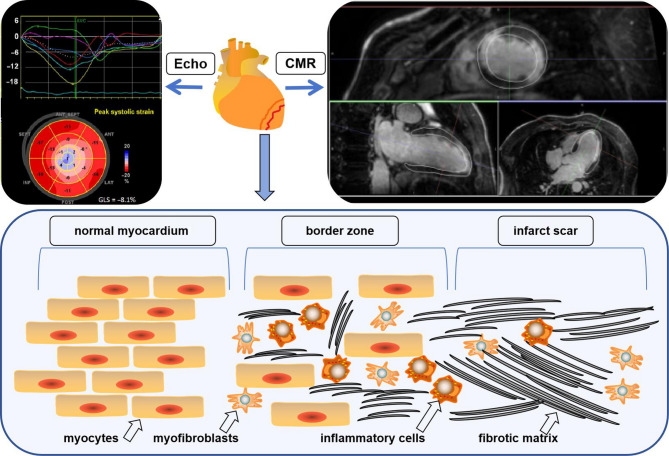
Ventricular arrhythmia (VA) is one of the common complications of many heart diseases in clinical practice, even some of its clinical symptoms are mild and non-specific, but the other may lead to sudden cardiac death (SCD) and most life-threatening VA is associated with ischemic cardiomyopathy (ICM). Nowadays, the developments in imaging techniques have provided clues to identify these highly variable VAs, which make clinicians identify patients with VA early and effectively who may have fatal consequences. Thereafter, it is beneficial to manage the risk stratification of patients, optimize their follow-up treatment, and improve clinical outcomes. This article reviews current ultrasound and magnetic resonance imaging techniques that can aid in diagnosis, treatment and prognosis, and provides clinicians with practical imaging and analytical recommendations to further identify patients with ICM who may develop VA. Clinical trial number Not applicable.
期刊介绍:
Echo Research and Practice aims to be the premier international journal for physicians, sonographers, nurses and other allied health professionals practising echocardiography and other cardiac imaging modalities. This open-access journal publishes quality clinical and basic research, reviews, videos, education materials and selected high-interest case reports and videos across all echocardiography modalities and disciplines, including paediatrics, anaesthetics, general practice, acute medicine and intensive care. Multi-modality studies primarily featuring the use of cardiac ultrasound in clinical practice, in association with Cardiac Computed Tomography, Cardiovascular Magnetic Resonance or Nuclear Cardiology are of interest. Topics include, but are not limited to: 2D echocardiography 3D echocardiography Comparative imaging techniques – CCT, CMR and Nuclear Cardiology Congenital heart disease, including foetal echocardiography Contrast echocardiography Critical care echocardiography Deformation imaging Doppler echocardiography Interventional echocardiography Intracardiac echocardiography Intraoperative echocardiography Prosthetic valves Stress echocardiography Technical innovations Transoesophageal echocardiography Valve disease.

 求助内容:
求助内容: 应助结果提醒方式:


