Viktor A Vilenskii, Maxim A Baushev, Leonid N Solomin
{"title":"上臂的参考线和角度。","authors":"Viktor A Vilenskii, Maxim A Baushev, Leonid N Solomin","doi":"10.5005/jp-journals-10080-1635","DOIUrl":null,"url":null,"abstract":"<p><strong>Aims and background: </strong>In the field of deformity analysis, the values for reference lines (both anatomical and mechanical) and reference angles of the femur and tibia are established. However, current data regarding the reference lines and angles of the humerus are limited, which limits comprehensive planning for deformity correction.The aim of this research was to establish standard values for the anatomical axis and angles of the humerus as measured in both the frontal and sagittal planes.</p><p><strong>Materials and methods: </strong>Radiographic images of the upper arms of 36 healthy participants (comprising 15 women and 21 men) were examined by utilizing two common imaging techniques: Anteroposterior and lateral views. Inclusion criteria for participants were over 18 years of age, have no prior upper limb injuries; no reports of pain in the upper limb joints; the lack of any musculoskeletal diseases and the absence of deformities. On the anteroposterior radiograph, assessments were made of joint intersections with the anatomical axis, along with the anatomical medial proximal humeral angle (aMPHA) and the anatomical lateral distal humeral angle (aLDHA). The lateral radiograph analysis focussed on joint intersections with the anatomical axis, the anatomical posterior proximal humerus angle (aPPHA) and the anatomical posterior distal humerus angle (aPDHA).</p><p><strong>Results: </strong>In the frontal plane, the anatomical axis intersected the proximal joint line of the humerus at the border of 36.6 ± 5.7 mm (76.57%) medially and 11.1 ± 4.5 mm (23.43%) laterally. At the distal joint line, the intersection occurred at the border of 22.5 ± 3.9 mm (37.88%) medially and 36.9 ± 5.6 mm (62.12%) laterally. In the sagittal plane, the anatomical axis intersected the proximal joint line at the border of 41.1 ± 11 mm (39.83%) in front and 62.1 ± 12.4 mm (60.17%) behind, and the distal joint line at the border of 16.1 ± 3.4 mm (76.3%) in front and 5.0 ± 2.1 mm (23.7%) behind. The following reference angle values were obtained: aMPHA = 45.2° ± 5.0°, aLDHA = 78° ± 4.1°, aPPHA = 56.8° ± 8.8° and aPDHA = 16.4° ± 3.1°.</p><p><strong>Conclusion: </strong>The obtained data will allow us to perform analysis, preoperative planning and evaluate the results of correction of humeral bone deformities with the accuracy required for clinical needs.</p><p><strong>Clinical significance: </strong>This study provides orthopaedic surgeons with new reference lines and angles of the humerus that are essential tools for deformity correction planning and estimating the results of deformity correction.</p><p><strong>How to cite this article: </strong>Vilenskii VA, Baushev MA, Solomin LN. Reference Lines and Angles of the Upper Arm. Strategies Trauma Limb Reconstr 2025;20(1):1-5.</p>","PeriodicalId":21979,"journal":{"name":"Strategies in Trauma and Limb Reconstruction","volume":"20 1","pages":"1-5"},"PeriodicalIF":1.3000,"publicationDate":"2025-01-01","publicationTypes":"Journal Article","fieldsOfStudy":null,"isOpenAccess":false,"openAccessPdf":"https://www.ncbi.nlm.nih.gov/pmc/articles/PMC12445131/pdf/","citationCount":"0","resultStr":"{\"title\":\"Reference Lines and Angles of the Upper Arm.\",\"authors\":\"Viktor A Vilenskii, Maxim A Baushev, Leonid N Solomin\",\"doi\":\"10.5005/jp-journals-10080-1635\",\"DOIUrl\":null,\"url\":null,\"abstract\":\"<p><strong>Aims and background: </strong>In the field of deformity analysis, the values for reference lines (both anatomical and mechanical) and reference angles of the femur and tibia are established. However, current data regarding the reference lines and angles of the humerus are limited, which limits comprehensive planning for deformity correction.The aim of this research was to establish standard values for the anatomical axis and angles of the humerus as measured in both the frontal and sagittal planes.</p><p><strong>Materials and methods: </strong>Radiographic images of the upper arms of 36 healthy participants (comprising 15 women and 21 men) were examined by utilizing two common imaging techniques: Anteroposterior and lateral views. Inclusion criteria for participants were over 18 years of age, have no prior upper limb injuries; no reports of pain in the upper limb joints; the lack of any musculoskeletal diseases and the absence of deformities. On the anteroposterior radiograph, assessments were made of joint intersections with the anatomical axis, along with the anatomical medial proximal humeral angle (aMPHA) and the anatomical lateral distal humeral angle (aLDHA). The lateral radiograph analysis focussed on joint intersections with the anatomical axis, the anatomical posterior proximal humerus angle (aPPHA) and the anatomical posterior distal humerus angle (aPDHA).</p><p><strong>Results: </strong>In the frontal plane, the anatomical axis intersected the proximal joint line of the humerus at the border of 36.6 ± 5.7 mm (76.57%) medially and 11.1 ± 4.5 mm (23.43%) laterally. At the distal joint line, the intersection occurred at the border of 22.5 ± 3.9 mm (37.88%) medially and 36.9 ± 5.6 mm (62.12%) laterally. In the sagittal plane, the anatomical axis intersected the proximal joint line at the border of 41.1 ± 11 mm (39.83%) in front and 62.1 ± 12.4 mm (60.17%) behind, and the distal joint line at the border of 16.1 ± 3.4 mm (76.3%) in front and 5.0 ± 2.1 mm (23.7%) behind. The following reference angle values were obtained: aMPHA = 45.2° ± 5.0°, aLDHA = 78° ± 4.1°, aPPHA = 56.8° ± 8.8° and aPDHA = 16.4° ± 3.1°.</p><p><strong>Conclusion: </strong>The obtained data will allow us to perform analysis, preoperative planning and evaluate the results of correction of humeral bone deformities with the accuracy required for clinical needs.</p><p><strong>Clinical significance: </strong>This study provides orthopaedic surgeons with new reference lines and angles of the humerus that are essential tools for deformity correction planning and estimating the results of deformity correction.</p><p><strong>How to cite this article: </strong>Vilenskii VA, Baushev MA, Solomin LN. Reference Lines and Angles of the Upper Arm. Strategies Trauma Limb Reconstr 2025;20(1):1-5.</p>\",\"PeriodicalId\":21979,\"journal\":{\"name\":\"Strategies in Trauma and Limb Reconstruction\",\"volume\":\"20 1\",\"pages\":\"1-5\"},\"PeriodicalIF\":1.3000,\"publicationDate\":\"2025-01-01\",\"publicationTypes\":\"Journal Article\",\"fieldsOfStudy\":null,\"isOpenAccess\":false,\"openAccessPdf\":\"https://www.ncbi.nlm.nih.gov/pmc/articles/PMC12445131/pdf/\",\"citationCount\":\"0\",\"resultStr\":null,\"platform\":\"Semanticscholar\",\"paperid\":null,\"PeriodicalName\":\"Strategies in Trauma and Limb Reconstruction\",\"FirstCategoryId\":\"1085\",\"ListUrlMain\":\"https://doi.org/10.5005/jp-journals-10080-1635\",\"RegionNum\":0,\"RegionCategory\":null,\"ArticlePicture\":[],\"TitleCN\":null,\"AbstractTextCN\":null,\"PMCID\":null,\"EPubDate\":\"2025/8/18 0:00:00\",\"PubModel\":\"Epub\",\"JCR\":\"Q3\",\"JCRName\":\"ORTHOPEDICS\",\"Score\":null,\"Total\":0}","platform":"Semanticscholar","paperid":null,"PeriodicalName":"Strategies in Trauma and Limb Reconstruction","FirstCategoryId":"1085","ListUrlMain":"https://doi.org/10.5005/jp-journals-10080-1635","RegionNum":0,"RegionCategory":null,"ArticlePicture":[],"TitleCN":null,"AbstractTextCN":null,"PMCID":null,"EPubDate":"2025/8/18 0:00:00","PubModel":"Epub","JCR":"Q3","JCRName":"ORTHOPEDICS","Score":null,"Total":0}
Aims and background: In the field of deformity analysis, the values for reference lines (both anatomical and mechanical) and reference angles of the femur and tibia are established. However, current data regarding the reference lines and angles of the humerus are limited, which limits comprehensive planning for deformity correction.The aim of this research was to establish standard values for the anatomical axis and angles of the humerus as measured in both the frontal and sagittal planes.
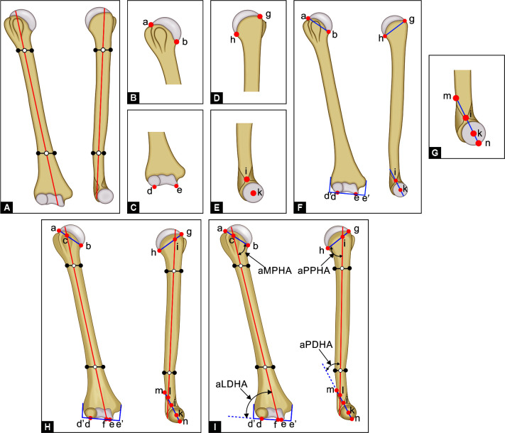
Materials and methods: Radiographic images of the upper arms of 36 healthy participants (comprising 15 women and 21 men) were examined by utilizing two common imaging techniques: Anteroposterior and lateral views. Inclusion criteria for participants were over 18 years of age, have no prior upper limb injuries; no reports of pain in the upper limb joints; the lack of any musculoskeletal diseases and the absence of deformities. On the anteroposterior radiograph, assessments were made of joint intersections with the anatomical axis, along with the anatomical medial proximal humeral angle (aMPHA) and the anatomical lateral distal humeral angle (aLDHA). The lateral radiograph analysis focussed on joint intersections with the anatomical axis, the anatomical posterior proximal humerus angle (aPPHA) and the anatomical posterior distal humerus angle (aPDHA).
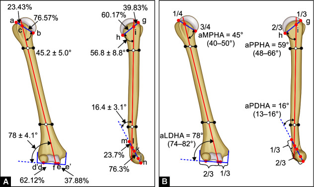
Results: In the frontal plane, the anatomical axis intersected the proximal joint line of the humerus at the border of 36.6 ± 5.7 mm (76.57%) medially and 11.1 ± 4.5 mm (23.43%) laterally. At the distal joint line, the intersection occurred at the border of 22.5 ± 3.9 mm (37.88%) medially and 36.9 ± 5.6 mm (62.12%) laterally. In the sagittal plane, the anatomical axis intersected the proximal joint line at the border of 41.1 ± 11 mm (39.83%) in front and 62.1 ± 12.4 mm (60.17%) behind, and the distal joint line at the border of 16.1 ± 3.4 mm (76.3%) in front and 5.0 ± 2.1 mm (23.7%) behind. The following reference angle values were obtained: aMPHA = 45.2° ± 5.0°, aLDHA = 78° ± 4.1°, aPPHA = 56.8° ± 8.8° and aPDHA = 16.4° ± 3.1°.
Conclusion: The obtained data will allow us to perform analysis, preoperative planning and evaluate the results of correction of humeral bone deformities with the accuracy required for clinical needs.
Clinical significance: This study provides orthopaedic surgeons with new reference lines and angles of the humerus that are essential tools for deformity correction planning and estimating the results of deformity correction.
How to cite this article: Vilenskii VA, Baushev MA, Solomin LN. Reference Lines and Angles of the Upper Arm. Strategies Trauma Limb Reconstr 2025;20(1):1-5.
期刊介绍:
Strategies in Trauma and Limb Reconstruction is dedicated to surgeons, allied medical professionals and researchers in the field of orthopaedics and trauma. The scope of the journal is to discuss the fields of skeletal injury, and the complications thereof, congenital and acquired limb deformities and deficiencies, and orthopaedic-related infection, together with their surgical and non-surgical treatments. The journal publishes original articles, reviews, case reports, descriptions of new or recognised treatment techniques, forum discussions of clinical scenarios and relevant correspondence. It aims to provide a widely accessible source of useful information to practitioners in the field through the problem- or technique-based approach of published articles.

 求助内容:
求助内容: 应助结果提醒方式:


