多参数肝脏MRI可解释病变分类的相关路径网络
IF 11.8
1区 医学
Q1 COMPUTER SCIENCE, ARTIFICIAL INTELLIGENCE
引用次数: 0
摘要
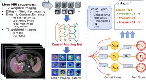
肝脏肿瘤的诊断是腹部影像学检查的关键任务,目前对局灶性肝脏病变(FLL)自动分类的研究较多。尽管有独特的诊断优势,但更多的是CT和超声,较少的是MRI。障碍在于肝脏MRI的数据集管理、技术复杂性和临床可解释性。在本文中,我们提出了相关路径网络(CRN),该网络采用10个MRI序列,预测病变类型(HCC、胆管瘤、转移、血管瘤、FNH、囊肿)和影像学特征,达到了较高的准确性和可解释性。CRN模型由编码分支、相关路由/中继模块和自关注模块组成。独立编码模式有助于信息解纠缠,相关路由方案有助于重定向和解耦,自关注增强了全局特征共享和预测一致性。该模型预测了详细的病变影像学特征,促进了可解释的分类和临床责任。我们还确定了信号关系并推导了定量的可解释性。我们的肝脏病变分类模型的良恶性准确率为97.2%,六类准确率为88%,平均影像特征准确率为84.9%,优于流行的CNN和基于变压器的模型。我们希望激发对多模态病变分类和模型可解释性的见解。本文章由计算机程序翻译,如有差异,请以英文原文为准。
Correlation Routing Network for Explainable Lesion Classification in Multi-Parametric Liver MRI
Liver tumor diagnosis is a key task in abdominal imaging examination, and there are numerous researches on automatic classifying focal liver lesions (FLL). More are in CT and ultrasound and fewer utilize MRI despite unique diagnostic advantages. The obstacles lie in dataset curation, technical complexity, and clinical explainability in liver MRI. In this paper, we propose the Correlation Routing Network (CRN) which takes in 10 MRI sequences and predicts lesion types (HCC, Cholangioma, Metastasis, Hemangioma, FNH, Cyst) as well as imaging features, to achieve both high accuracy and explainability. The CRN model consists of encoding branches, correlation routing/relay modules, and the self-attention module. The independent encoding paradigm facilitates information disentangling, the correlation routing scheme helps redirection and decoupling effectively, and the self-attention enforces global feature sharing and prediction consistency. The model predicts detailed lesion imaging features, promoting explainable classification and clinical accountability. We also identify the signal relations and derive quantitative explainability. Our liver lesion classification model achieves malignant-benign accuracy of 97.2%, six-class accuracy of 88%, and averaged imaging feature accuracy of 84.9%, outperforming popular CNN and transformer-based models. We hope to spark insights for multimodal lesion classification and model explainability.
求助全文
通过发布文献求助,成功后即可免费获取论文全文。
去求助
来源期刊

Medical image analysis
工程技术-工程:生物医学
CiteScore
22.10
自引率
6.40%
发文量
309
审稿时长
6.6 months
期刊介绍:
Medical Image Analysis serves as a platform for sharing new research findings in the realm of medical and biological image analysis, with a focus on applications of computer vision, virtual reality, and robotics to biomedical imaging challenges. The journal prioritizes the publication of high-quality, original papers contributing to the fundamental science of processing, analyzing, and utilizing medical and biological images. It welcomes approaches utilizing biomedical image datasets across all spatial scales, from molecular/cellular imaging to tissue/organ imaging.
 求助内容:
求助内容: 应助结果提醒方式:


