Anaam R Alhadeethi, Steffy Terrance, Mohamed E Hassan, Khalid Al Ali
{"title":"巨大间皮性脾囊肿。","authors":"Anaam R Alhadeethi, Steffy Terrance, Mohamed E Hassan, Khalid Al Ali","doi":"10.4293/CRSLS.2025.00046","DOIUrl":null,"url":null,"abstract":"<p><strong>Introduction: </strong>Splenic cysts are rare lesions that are classified as either true (primary) or false (secondary) cysts based on their epithelial lining. The pathogenesis of primary splenic cysts is not well understood, and several hypotheses have been proposed, including the Mesothelial invagination theory, which postulates that during development, the mesothelial lining invades along with the capsule. As the lining has a pluripotent nature, it has the propensity to undergo metaplasia and secretion of fluid, leading to the formation of cysts.</p><p><strong>Case presentation: </strong>A 12-year-old female patient presented with a visible upper abdominal, painless cystic lesion, underwent blood tests and radiological diagnostic tools, such as abdominal ultrasound and computed tomography (CT) scan, but no definite diagnosis could be reached. Ultrasound-guided aspiration of the cyst was done, followed by explorative laparoscopy with total excision of the cyst (which was found to originate from the spleen), accompanied by partial splenectomy. The cyst was diagnosed as a benign primary mesothelial cyst of the spleen by histopathology. The patient experienced an uneventful postoperative period and showed no recurrence during follow-up.</p><p><strong>Conclusion: </strong>A significant challenge for surgeons in terms of diagnosis, surgical planning, and managing intraoperative surprises is the difficulty in detecting the origin and nature of a large abdominal cyst, despite the availability of highly sophisticated diagnostic tools. Minimally invasive partial splenectomy in the pediatric age group is a feasible surgical intervention.</p>","PeriodicalId":72723,"journal":{"name":"CRSLS : MIS case reports from SLS","volume":"12 3","pages":""},"PeriodicalIF":0.0000,"publicationDate":"2025-09-17","publicationTypes":"Journal Article","fieldsOfStudy":null,"isOpenAccess":false,"openAccessPdf":"https://www.ncbi.nlm.nih.gov/pmc/articles/PMC12442189/pdf/","citationCount":"0","resultStr":"{\"title\":\"Huge Mesothelial Splenic Cyst.\",\"authors\":\"Anaam R Alhadeethi, Steffy Terrance, Mohamed E Hassan, Khalid Al Ali\",\"doi\":\"10.4293/CRSLS.2025.00046\",\"DOIUrl\":null,\"url\":null,\"abstract\":\"<p><strong>Introduction: </strong>Splenic cysts are rare lesions that are classified as either true (primary) or false (secondary) cysts based on their epithelial lining. The pathogenesis of primary splenic cysts is not well understood, and several hypotheses have been proposed, including the Mesothelial invagination theory, which postulates that during development, the mesothelial lining invades along with the capsule. As the lining has a pluripotent nature, it has the propensity to undergo metaplasia and secretion of fluid, leading to the formation of cysts.</p><p><strong>Case presentation: </strong>A 12-year-old female patient presented with a visible upper abdominal, painless cystic lesion, underwent blood tests and radiological diagnostic tools, such as abdominal ultrasound and computed tomography (CT) scan, but no definite diagnosis could be reached. Ultrasound-guided aspiration of the cyst was done, followed by explorative laparoscopy with total excision of the cyst (which was found to originate from the spleen), accompanied by partial splenectomy. The cyst was diagnosed as a benign primary mesothelial cyst of the spleen by histopathology. The patient experienced an uneventful postoperative period and showed no recurrence during follow-up.</p><p><strong>Conclusion: </strong>A significant challenge for surgeons in terms of diagnosis, surgical planning, and managing intraoperative surprises is the difficulty in detecting the origin and nature of a large abdominal cyst, despite the availability of highly sophisticated diagnostic tools. Minimally invasive partial splenectomy in the pediatric age group is a feasible surgical intervention.</p>\",\"PeriodicalId\":72723,\"journal\":{\"name\":\"CRSLS : MIS case reports from SLS\",\"volume\":\"12 3\",\"pages\":\"\"},\"PeriodicalIF\":0.0000,\"publicationDate\":\"2025-09-17\",\"publicationTypes\":\"Journal Article\",\"fieldsOfStudy\":null,\"isOpenAccess\":false,\"openAccessPdf\":\"https://www.ncbi.nlm.nih.gov/pmc/articles/PMC12442189/pdf/\",\"citationCount\":\"0\",\"resultStr\":null,\"platform\":\"Semanticscholar\",\"paperid\":null,\"PeriodicalName\":\"CRSLS : MIS case reports from SLS\",\"FirstCategoryId\":\"1085\",\"ListUrlMain\":\"https://doi.org/10.4293/CRSLS.2025.00046\",\"RegionNum\":0,\"RegionCategory\":null,\"ArticlePicture\":[],\"TitleCN\":null,\"AbstractTextCN\":null,\"PMCID\":null,\"EPubDate\":\"2025/7/1 0:00:00\",\"PubModel\":\"eCollection\",\"JCR\":\"\",\"JCRName\":\"\",\"Score\":null,\"Total\":0}","platform":"Semanticscholar","paperid":null,"PeriodicalName":"CRSLS : MIS case reports from SLS","FirstCategoryId":"1085","ListUrlMain":"https://doi.org/10.4293/CRSLS.2025.00046","RegionNum":0,"RegionCategory":null,"ArticlePicture":[],"TitleCN":null,"AbstractTextCN":null,"PMCID":null,"EPubDate":"2025/7/1 0:00:00","PubModel":"eCollection","JCR":"","JCRName":"","Score":null,"Total":0}
Introduction: Splenic cysts are rare lesions that are classified as either true (primary) or false (secondary) cysts based on their epithelial lining. The pathogenesis of primary splenic cysts is not well understood, and several hypotheses have been proposed, including the Mesothelial invagination theory, which postulates that during development, the mesothelial lining invades along with the capsule. As the lining has a pluripotent nature, it has the propensity to undergo metaplasia and secretion of fluid, leading to the formation of cysts.
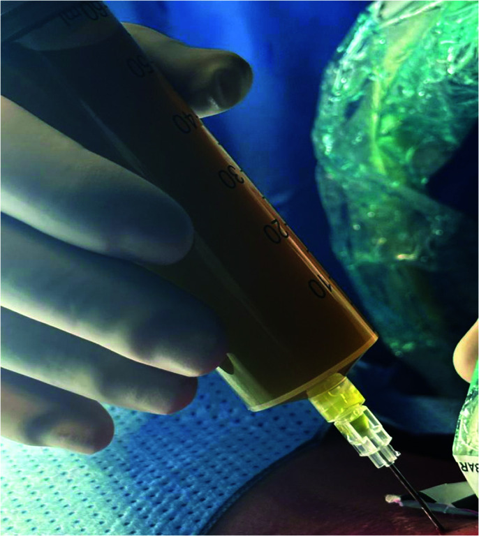
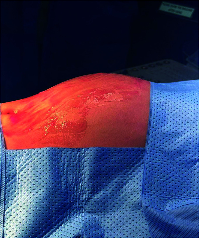
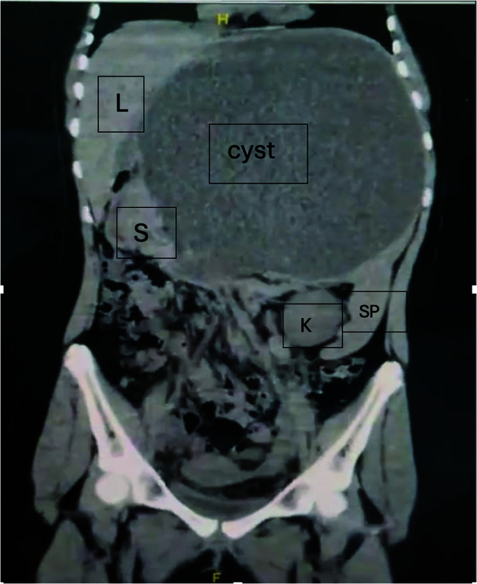
Case presentation: A 12-year-old female patient presented with a visible upper abdominal, painless cystic lesion, underwent blood tests and radiological diagnostic tools, such as abdominal ultrasound and computed tomography (CT) scan, but no definite diagnosis could be reached. Ultrasound-guided aspiration of the cyst was done, followed by explorative laparoscopy with total excision of the cyst (which was found to originate from the spleen), accompanied by partial splenectomy. The cyst was diagnosed as a benign primary mesothelial cyst of the spleen by histopathology. The patient experienced an uneventful postoperative period and showed no recurrence during follow-up.
Conclusion: A significant challenge for surgeons in terms of diagnosis, surgical planning, and managing intraoperative surprises is the difficulty in detecting the origin and nature of a large abdominal cyst, despite the availability of highly sophisticated diagnostic tools. Minimally invasive partial splenectomy in the pediatric age group is a feasible surgical intervention.

 求助内容:
求助内容: 应助结果提醒方式:


