M Forooghi, A Askari, M Haghdel, A G Haghighi, M H Anbardar, A H Hassani, H Foroutan, A S Aloudal, Sh Yousufzai
{"title":"工程膀胱修复的未来:生物相容性3d打印支架能否作为肠段治疗膀胱外翻的新替代品?","authors":"M Forooghi, A Askari, M Haghdel, A G Haghighi, M H Anbardar, A H Hassani, H Foroutan, A S Aloudal, Sh Yousufzai","doi":"10.1155/aiu/9437696","DOIUrl":null,"url":null,"abstract":"<p><p><b>Background:</b> Bladder reconstruction traditionally involves intestinal segments, which, despite their effectiveness, carry significant risks such as metabolic disturbances and infection. Safer, synthetic alternatives are needed. We evaluated a novel 3D-printed multilayered bladder scaffold combining polylactic acid (PLA), thermoplastic polyurethane (TPU), and polyvinyl alcohol (PVA) in a rabbit model. <b>Methods:</b> Anatomically tailored scaffolds were designed using computer-aided design (CAD) and fabricated under good manufacturing practice (GMP) conditions. Mechanical integrity was assessed after 60 days of incubation in simulated bladder media, including measurements of modulus of elasticity, tensile strength, elongation, and shape recovery. Acid/alkaline resistance was tested for chemical stability. For in vivo analysis, four rabbits underwent bladder augmentation with a 1 × 1 cm scaffold-augmented defect. Postoperative outcomes were monitored for 60 days, followed by histopathological evaluation. <b>Results:</b> After incubation, the scaffolds retained mechanical strength (modulus: 1.2 ± 0.3 GPa; tensile strength: 18.5 ± 2.1 MPa) with minimal elongation reduction (25% vs. 28% unused). Chemical testing confirmed structural stability and full shape recovery. In vivo, all rabbits survived without urinary leakage. Mild intra-abdominal adhesions and universal cystolithiasis were noted. Histology showed complete urothelial reepithelialization and mild-to-moderate submucosal fibrosis with chronic inflammation but no necrosis or acute inflammation. Compared to biological scaffolds, the synthetic construct showed reduced mortality and comparable inflammation, though with increased stone formation. <b>Conclusion:</b> This 3D-printed scaffold demonstrates promising biocompatibility, mechanical durability, and integration in bladder repair. While early results are encouraging, further studies with larger sample sizes and longer follow-up are needed to address limitations such as cystolithiasis risk.</p>","PeriodicalId":7490,"journal":{"name":"Advances in Urology","volume":"2025 ","pages":"9437696"},"PeriodicalIF":2.3000,"publicationDate":"2025-09-10","publicationTypes":"Journal Article","fieldsOfStudy":null,"isOpenAccess":false,"openAccessPdf":"https://www.ncbi.nlm.nih.gov/pmc/articles/PMC12443514/pdf/","citationCount":"0","resultStr":"{\"title\":\"Engineering the Future of Bladder Repair: Can Biocompatible 3D-Printed Scaffolds Serve as a Novel Alternative to Intestinal Segments for the Treatment of Bladder Exstrophy?\",\"authors\":\"M Forooghi, A Askari, M Haghdel, A G Haghighi, M H Anbardar, A H Hassani, H Foroutan, A S Aloudal, Sh Yousufzai\",\"doi\":\"10.1155/aiu/9437696\",\"DOIUrl\":null,\"url\":null,\"abstract\":\"<p><p><b>Background:</b> Bladder reconstruction traditionally involves intestinal segments, which, despite their effectiveness, carry significant risks such as metabolic disturbances and infection. Safer, synthetic alternatives are needed. We evaluated a novel 3D-printed multilayered bladder scaffold combining polylactic acid (PLA), thermoplastic polyurethane (TPU), and polyvinyl alcohol (PVA) in a rabbit model. <b>Methods:</b> Anatomically tailored scaffolds were designed using computer-aided design (CAD) and fabricated under good manufacturing practice (GMP) conditions. Mechanical integrity was assessed after 60 days of incubation in simulated bladder media, including measurements of modulus of elasticity, tensile strength, elongation, and shape recovery. Acid/alkaline resistance was tested for chemical stability. For in vivo analysis, four rabbits underwent bladder augmentation with a 1 × 1 cm scaffold-augmented defect. Postoperative outcomes were monitored for 60 days, followed by histopathological evaluation. <b>Results:</b> After incubation, the scaffolds retained mechanical strength (modulus: 1.2 ± 0.3 GPa; tensile strength: 18.5 ± 2.1 MPa) with minimal elongation reduction (25% vs. 28% unused). Chemical testing confirmed structural stability and full shape recovery. In vivo, all rabbits survived without urinary leakage. Mild intra-abdominal adhesions and universal cystolithiasis were noted. Histology showed complete urothelial reepithelialization and mild-to-moderate submucosal fibrosis with chronic inflammation but no necrosis or acute inflammation. Compared to biological scaffolds, the synthetic construct showed reduced mortality and comparable inflammation, though with increased stone formation. <b>Conclusion:</b> This 3D-printed scaffold demonstrates promising biocompatibility, mechanical durability, and integration in bladder repair. While early results are encouraging, further studies with larger sample sizes and longer follow-up are needed to address limitations such as cystolithiasis risk.</p>\",\"PeriodicalId\":7490,\"journal\":{\"name\":\"Advances in Urology\",\"volume\":\"2025 \",\"pages\":\"9437696\"},\"PeriodicalIF\":2.3000,\"publicationDate\":\"2025-09-10\",\"publicationTypes\":\"Journal Article\",\"fieldsOfStudy\":null,\"isOpenAccess\":false,\"openAccessPdf\":\"https://www.ncbi.nlm.nih.gov/pmc/articles/PMC12443514/pdf/\",\"citationCount\":\"0\",\"resultStr\":null,\"platform\":\"Semanticscholar\",\"paperid\":null,\"PeriodicalName\":\"Advances in Urology\",\"FirstCategoryId\":\"1085\",\"ListUrlMain\":\"https://doi.org/10.1155/aiu/9437696\",\"RegionNum\":0,\"RegionCategory\":null,\"ArticlePicture\":[],\"TitleCN\":null,\"AbstractTextCN\":null,\"PMCID\":null,\"EPubDate\":\"2025/1/1 0:00:00\",\"PubModel\":\"eCollection\",\"JCR\":\"Q3\",\"JCRName\":\"UROLOGY & NEPHROLOGY\",\"Score\":null,\"Total\":0}","platform":"Semanticscholar","paperid":null,"PeriodicalName":"Advances in Urology","FirstCategoryId":"1085","ListUrlMain":"https://doi.org/10.1155/aiu/9437696","RegionNum":0,"RegionCategory":null,"ArticlePicture":[],"TitleCN":null,"AbstractTextCN":null,"PMCID":null,"EPubDate":"2025/1/1 0:00:00","PubModel":"eCollection","JCR":"Q3","JCRName":"UROLOGY & NEPHROLOGY","Score":null,"Total":0}
引用次数: 0
摘要
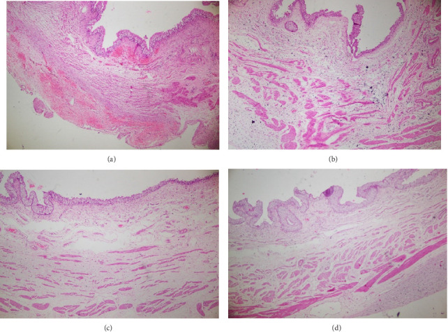
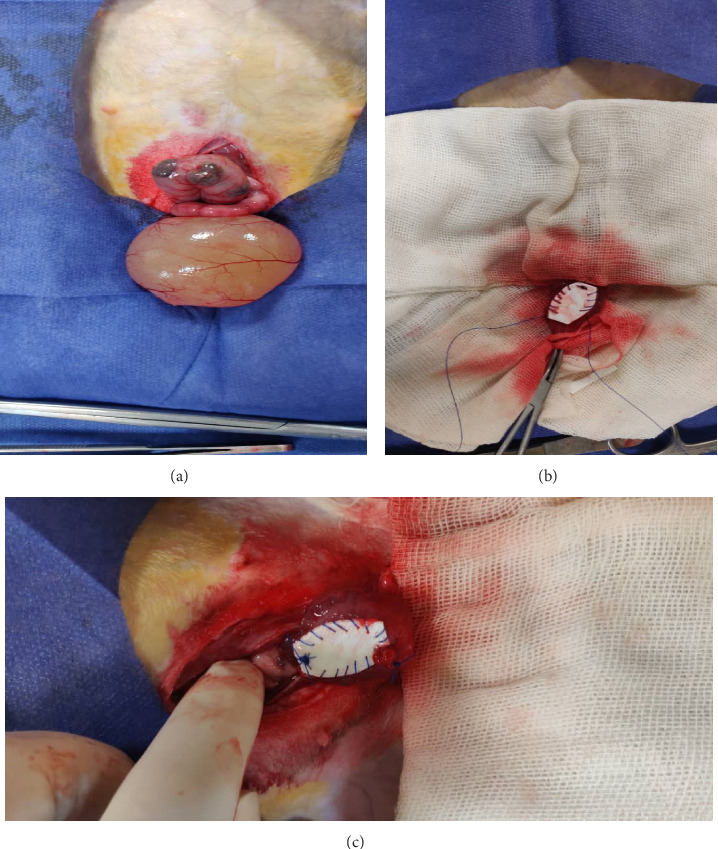
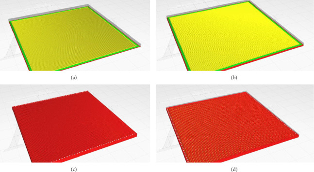
背景:膀胱重建传统上涉及肠段,尽管其有效,但存在代谢紊乱和感染等重大风险。需要更安全的合成替代品。我们在兔模型中评估了一种新型3d打印多层膀胱支架,该支架由聚乳酸(PLA)、热塑性聚氨酯(TPU)和聚乙烯醇(PVA)组成。方法:采用计算机辅助设计(CAD)设计解剖定制支架,并在GMP条件下制作。在模拟膀胱介质中培养60天后评估机械完整性,包括弹性模量、拉伸强度、伸长率和形状恢复的测量。对其耐酸碱性进行了化学稳定性测试。在体内分析中,4只兔子接受了膀胱增强术,伴有1 × 1厘米的支架增强缺陷。术后监测60天,然后进行组织病理学评估。结果:培养后,支架保持机械强度(模量:1.2±0.3 GPa;抗拉强度:18.5±2.1 MPa),延伸率最小(25% vs 28%未使用)。化学测试证实了结构的稳定性和完全的形状恢复。在体内,所有家兔均存活,无尿漏。轻度腹内粘连和普遍膀胱结石。组织学表现为完全的尿路上皮再上皮化和轻至中度粘膜下纤维化,伴慢性炎症,但无坏死或急性炎症。与生物支架相比,合成支架显示出较低的死亡率和类似的炎症,尽管会增加结石的形成。结论:该3d打印支架具有良好的生物相容性、机械耐久性和膀胱修复的整体性。虽然早期结果令人鼓舞,但需要进一步的研究,样本量更大,随访时间更长,以解决诸如膀胱结石风险等局限性。
Engineering the Future of Bladder Repair: Can Biocompatible 3D-Printed Scaffolds Serve as a Novel Alternative to Intestinal Segments for the Treatment of Bladder Exstrophy?
Background: Bladder reconstruction traditionally involves intestinal segments, which, despite their effectiveness, carry significant risks such as metabolic disturbances and infection. Safer, synthetic alternatives are needed. We evaluated a novel 3D-printed multilayered bladder scaffold combining polylactic acid (PLA), thermoplastic polyurethane (TPU), and polyvinyl alcohol (PVA) in a rabbit model. Methods: Anatomically tailored scaffolds were designed using computer-aided design (CAD) and fabricated under good manufacturing practice (GMP) conditions. Mechanical integrity was assessed after 60 days of incubation in simulated bladder media, including measurements of modulus of elasticity, tensile strength, elongation, and shape recovery. Acid/alkaline resistance was tested for chemical stability. For in vivo analysis, four rabbits underwent bladder augmentation with a 1 × 1 cm scaffold-augmented defect. Postoperative outcomes were monitored for 60 days, followed by histopathological evaluation. Results: After incubation, the scaffolds retained mechanical strength (modulus: 1.2 ± 0.3 GPa; tensile strength: 18.5 ± 2.1 MPa) with minimal elongation reduction (25% vs. 28% unused). Chemical testing confirmed structural stability and full shape recovery. In vivo, all rabbits survived without urinary leakage. Mild intra-abdominal adhesions and universal cystolithiasis were noted. Histology showed complete urothelial reepithelialization and mild-to-moderate submucosal fibrosis with chronic inflammation but no necrosis or acute inflammation. Compared to biological scaffolds, the synthetic construct showed reduced mortality and comparable inflammation, though with increased stone formation. Conclusion: This 3D-printed scaffold demonstrates promising biocompatibility, mechanical durability, and integration in bladder repair. While early results are encouraging, further studies with larger sample sizes and longer follow-up are needed to address limitations such as cystolithiasis risk.
期刊介绍:
Advances in Urology is a peer-reviewed, open access journal that publishes state-of-the-art reviews and original research papers of wide interest in all fields of urology. The journal strives to provide publication of important manuscripts to the widest possible audience worldwide, without the constraints of expensive, hard-to-access, traditional bound journals. Advances in Urology is designed to improve publication access of both well-established urologic scientists and less well-established writers, by allowing interested scientists worldwide to participate fully.

 求助内容:
求助内容: 应助结果提醒方式:


