Clara Massaguer, Irene De Haro Jorge, Laura Saura García, Jordi Prat Ortells, María Elena Muñoz Fernández, Xavier Tarrado
{"title":"消失的胃裂:在一个看似正常的腹部产前诊断的重要性。","authors":"Clara Massaguer, Irene De Haro Jorge, Laura Saura García, Jordi Prat Ortells, María Elena Muñoz Fernández, Xavier Tarrado","doi":"10.1055/a-2692-6661","DOIUrl":null,"url":null,"abstract":"<p><p>A newborn of 32 + 6 weeks' gestational age with prenatal diagnosis of gastroschisis was born through elective caesarean section. Ultrasonography at 16 + 4 gestational weeks (GW) showed a gastroschisis with free bowel loops floating in amniotic fluid. From 27 + 4 GW onward, serial ultrasounds showed the disappearance of extra-abdominal intestine and progressive intra-abdominal intestinal loops dilation, raising suspicion for vanishing gastroschisis. Birth weight was 2,136 grams and the external appearance of the abdomen was normal. An exploratory laparotomy was performed, finding a dilated proximal jejunal loop with a type III intestinal atresia, microcolon, and no other remainder bowel in between. The total length of the small intestine was 21 cm. Serial transverse enteroplasties for intestinal lengthening (reaching 38 cm), along with lateroterminal jejunocolic anastomosis were performed. The patient was discharged after 5 months of hospitalization with home parenteral nutrition. At 2 years and 8 months of age, the child is thriving and off parenteral support. Vanishing gastroschisis is a rare and severe form of complex gastroschisis whose prenatal diagnosis is crucial for parental counseling, timely delivery, and early surgical intervention. Multidisciplinary approach is essential to manage intestinal failure and improve long-term outcomes in these patients.</p>","PeriodicalId":43204,"journal":{"name":"European Journal of Pediatric Surgery Reports","volume":"13 1","pages":"e151-e154"},"PeriodicalIF":0.7000,"publicationDate":"2025-09-12","publicationTypes":"Journal Article","fieldsOfStudy":null,"isOpenAccess":false,"openAccessPdf":"https://www.ncbi.nlm.nih.gov/pmc/articles/PMC12431809/pdf/","citationCount":"0","resultStr":"{\"title\":\"Vanishing Gastroschisis: The Importance of Prenatal Diagnosis in a Seemingly Normal Abdomen.\",\"authors\":\"Clara Massaguer, Irene De Haro Jorge, Laura Saura García, Jordi Prat Ortells, María Elena Muñoz Fernández, Xavier Tarrado\",\"doi\":\"10.1055/a-2692-6661\",\"DOIUrl\":null,\"url\":null,\"abstract\":\"<p><p>A newborn of 32 + 6 weeks' gestational age with prenatal diagnosis of gastroschisis was born through elective caesarean section. Ultrasonography at 16 + 4 gestational weeks (GW) showed a gastroschisis with free bowel loops floating in amniotic fluid. From 27 + 4 GW onward, serial ultrasounds showed the disappearance of extra-abdominal intestine and progressive intra-abdominal intestinal loops dilation, raising suspicion for vanishing gastroschisis. Birth weight was 2,136 grams and the external appearance of the abdomen was normal. An exploratory laparotomy was performed, finding a dilated proximal jejunal loop with a type III intestinal atresia, microcolon, and no other remainder bowel in between. The total length of the small intestine was 21 cm. Serial transverse enteroplasties for intestinal lengthening (reaching 38 cm), along with lateroterminal jejunocolic anastomosis were performed. The patient was discharged after 5 months of hospitalization with home parenteral nutrition. At 2 years and 8 months of age, the child is thriving and off parenteral support. Vanishing gastroschisis is a rare and severe form of complex gastroschisis whose prenatal diagnosis is crucial for parental counseling, timely delivery, and early surgical intervention. Multidisciplinary approach is essential to manage intestinal failure and improve long-term outcomes in these patients.</p>\",\"PeriodicalId\":43204,\"journal\":{\"name\":\"European Journal of Pediatric Surgery Reports\",\"volume\":\"13 1\",\"pages\":\"e151-e154\"},\"PeriodicalIF\":0.7000,\"publicationDate\":\"2025-09-12\",\"publicationTypes\":\"Journal Article\",\"fieldsOfStudy\":null,\"isOpenAccess\":false,\"openAccessPdf\":\"https://www.ncbi.nlm.nih.gov/pmc/articles/PMC12431809/pdf/\",\"citationCount\":\"0\",\"resultStr\":null,\"platform\":\"Semanticscholar\",\"paperid\":null,\"PeriodicalName\":\"European Journal of Pediatric Surgery Reports\",\"FirstCategoryId\":\"1085\",\"ListUrlMain\":\"https://doi.org/10.1055/a-2692-6661\",\"RegionNum\":0,\"RegionCategory\":null,\"ArticlePicture\":[],\"TitleCN\":null,\"AbstractTextCN\":null,\"PMCID\":null,\"EPubDate\":\"2025/1/1 0:00:00\",\"PubModel\":\"eCollection\",\"JCR\":\"Q4\",\"JCRName\":\"SURGERY\",\"Score\":null,\"Total\":0}","platform":"Semanticscholar","paperid":null,"PeriodicalName":"European Journal of Pediatric Surgery Reports","FirstCategoryId":"1085","ListUrlMain":"https://doi.org/10.1055/a-2692-6661","RegionNum":0,"RegionCategory":null,"ArticlePicture":[],"TitleCN":null,"AbstractTextCN":null,"PMCID":null,"EPubDate":"2025/1/1 0:00:00","PubModel":"eCollection","JCR":"Q4","JCRName":"SURGERY","Score":null,"Total":0}
Vanishing Gastroschisis: The Importance of Prenatal Diagnosis in a Seemingly Normal Abdomen.
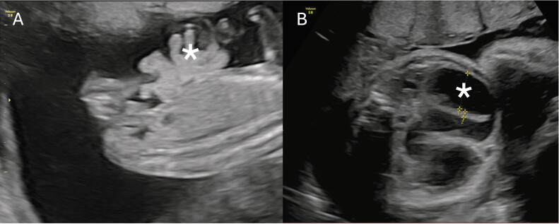
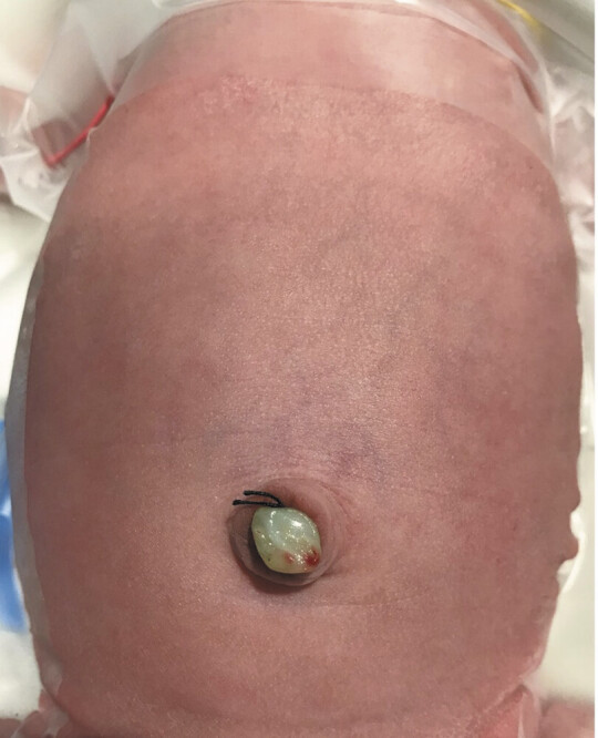
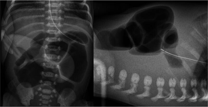
A newborn of 32 + 6 weeks' gestational age with prenatal diagnosis of gastroschisis was born through elective caesarean section. Ultrasonography at 16 + 4 gestational weeks (GW) showed a gastroschisis with free bowel loops floating in amniotic fluid. From 27 + 4 GW onward, serial ultrasounds showed the disappearance of extra-abdominal intestine and progressive intra-abdominal intestinal loops dilation, raising suspicion for vanishing gastroschisis. Birth weight was 2,136 grams and the external appearance of the abdomen was normal. An exploratory laparotomy was performed, finding a dilated proximal jejunal loop with a type III intestinal atresia, microcolon, and no other remainder bowel in between. The total length of the small intestine was 21 cm. Serial transverse enteroplasties for intestinal lengthening (reaching 38 cm), along with lateroterminal jejunocolic anastomosis were performed. The patient was discharged after 5 months of hospitalization with home parenteral nutrition. At 2 years and 8 months of age, the child is thriving and off parenteral support. Vanishing gastroschisis is a rare and severe form of complex gastroschisis whose prenatal diagnosis is crucial for parental counseling, timely delivery, and early surgical intervention. Multidisciplinary approach is essential to manage intestinal failure and improve long-term outcomes in these patients.

 求助内容:
求助内容: 应助结果提醒方式:


