{"title":"胸椎与L1 CT衰减预测骨质疏松的比较。","authors":"Lilan Wu, Shunfa Huang, Liling Xu, Shengxiang Rao, Zhen Qian, Mengze Zhang, Ying Yuan, Jianjun Zhou","doi":"10.1177/1759720X251374134","DOIUrl":null,"url":null,"abstract":"<p><strong>Background: </strong>Since dual-energy x-ray absorptiometry (DXA) is currently the most commonly used reference standard, most previous studies using computed tomography (CT) attenuation values to predict osteoporosis have chosen abdominal CT images. A few studies have investigated whether the thoracic vertebrae can be independently used for the identification of osteoporosis compared to the lumbar vertebrae.</p><p><strong>Objective: </strong>To investigate whether the attenuation values of thoracic vertebrae measured using artificial intelligence (AI) on chest CT would independently predict osteoporosis identification, considering central DXA as a reference standard.</p><p><strong>Design: </strong>This was a cross-sectional study.</p><p><strong>Methods: </strong>A total of 553 participants (353 men and 200 women) who underwent chest CT and DXA within 1 day were included. The attenuation values (HU) of the T7-12 vertebrae and L1 vertebra were obtained by AI. The effects of the clinical baseline data and attenuation values among the normal, osteopenia, and osteoporosis groups were compared. The correlation between attenuation and bone mineral density (BMD) values was analyzed, and the diagnostic performance of thoracic and first lumbar vertebrae attenuation values for diagnosing osteopenia or osteoporosis was further explored.</p><p><strong>Results: </strong>The CT attenuation values of T7-12 and L1 vertebrae showed positive correlation with <i>T</i>-score (<i>R</i> = 0.58-0.61, <i>p</i> < 0.01). T12 attenuation >184.8 HU was 84.1% sensitive and 70.6% specific for distinguishing normal BMD, while T12 attenuation <146.2 HU was 61.4% specific and 75.6% sensitive for distinguishing osteoporosis from osteopenia. There were no significant differences between the T10-12 and L1 groups in distinguishing the normal, osteopenia, and osteoporosis groups. Moreover, the diagnostic efficacy among the T10, T11, T12, and L1 vertebral bodies was not statistically significantly different among the three groups.</p><p><strong>Conclusion: </strong>Opportunistic screening is a valid method for predicting osteopenia or osteoporosis. As a rapid and effective tool, T10-12 vertebral attenuation measures can be incorporated to predict osteoporosis and identify patients who may benefit from further investigations using DXA based on routine clinical chest CT examinations.</p>","PeriodicalId":23056,"journal":{"name":"Therapeutic Advances in Musculoskeletal Disease","volume":"17 ","pages":"1759720X251374134"},"PeriodicalIF":4.1000,"publicationDate":"2025-09-11","publicationTypes":"Journal Article","fieldsOfStudy":null,"isOpenAccess":false,"openAccessPdf":"https://www.ncbi.nlm.nih.gov/pmc/articles/PMC12432313/pdf/","citationCount":"0","resultStr":"{\"title\":\"Comparison of thoracic vertebrae and L1 CT attenuation in predicting osteoporosis using opportunistic chest CT.\",\"authors\":\"Lilan Wu, Shunfa Huang, Liling Xu, Shengxiang Rao, Zhen Qian, Mengze Zhang, Ying Yuan, Jianjun Zhou\",\"doi\":\"10.1177/1759720X251374134\",\"DOIUrl\":null,\"url\":null,\"abstract\":\"<p><strong>Background: </strong>Since dual-energy x-ray absorptiometry (DXA) is currently the most commonly used reference standard, most previous studies using computed tomography (CT) attenuation values to predict osteoporosis have chosen abdominal CT images. A few studies have investigated whether the thoracic vertebrae can be independently used for the identification of osteoporosis compared to the lumbar vertebrae.</p><p><strong>Objective: </strong>To investigate whether the attenuation values of thoracic vertebrae measured using artificial intelligence (AI) on chest CT would independently predict osteoporosis identification, considering central DXA as a reference standard.</p><p><strong>Design: </strong>This was a cross-sectional study.</p><p><strong>Methods: </strong>A total of 553 participants (353 men and 200 women) who underwent chest CT and DXA within 1 day were included. The attenuation values (HU) of the T7-12 vertebrae and L1 vertebra were obtained by AI. The effects of the clinical baseline data and attenuation values among the normal, osteopenia, and osteoporosis groups were compared. The correlation between attenuation and bone mineral density (BMD) values was analyzed, and the diagnostic performance of thoracic and first lumbar vertebrae attenuation values for diagnosing osteopenia or osteoporosis was further explored.</p><p><strong>Results: </strong>The CT attenuation values of T7-12 and L1 vertebrae showed positive correlation with <i>T</i>-score (<i>R</i> = 0.58-0.61, <i>p</i> < 0.01). T12 attenuation >184.8 HU was 84.1% sensitive and 70.6% specific for distinguishing normal BMD, while T12 attenuation <146.2 HU was 61.4% specific and 75.6% sensitive for distinguishing osteoporosis from osteopenia. There were no significant differences between the T10-12 and L1 groups in distinguishing the normal, osteopenia, and osteoporosis groups. Moreover, the diagnostic efficacy among the T10, T11, T12, and L1 vertebral bodies was not statistically significantly different among the three groups.</p><p><strong>Conclusion: </strong>Opportunistic screening is a valid method for predicting osteopenia or osteoporosis. As a rapid and effective tool, T10-12 vertebral attenuation measures can be incorporated to predict osteoporosis and identify patients who may benefit from further investigations using DXA based on routine clinical chest CT examinations.</p>\",\"PeriodicalId\":23056,\"journal\":{\"name\":\"Therapeutic Advances in Musculoskeletal Disease\",\"volume\":\"17 \",\"pages\":\"1759720X251374134\"},\"PeriodicalIF\":4.1000,\"publicationDate\":\"2025-09-11\",\"publicationTypes\":\"Journal Article\",\"fieldsOfStudy\":null,\"isOpenAccess\":false,\"openAccessPdf\":\"https://www.ncbi.nlm.nih.gov/pmc/articles/PMC12432313/pdf/\",\"citationCount\":\"0\",\"resultStr\":null,\"platform\":\"Semanticscholar\",\"paperid\":null,\"PeriodicalName\":\"Therapeutic Advances in Musculoskeletal Disease\",\"FirstCategoryId\":\"3\",\"ListUrlMain\":\"https://doi.org/10.1177/1759720X251374134\",\"RegionNum\":2,\"RegionCategory\":\"医学\",\"ArticlePicture\":[],\"TitleCN\":null,\"AbstractTextCN\":null,\"PMCID\":null,\"EPubDate\":\"2025/1/1 0:00:00\",\"PubModel\":\"eCollection\",\"JCR\":\"Q2\",\"JCRName\":\"RHEUMATOLOGY\",\"Score\":null,\"Total\":0}","platform":"Semanticscholar","paperid":null,"PeriodicalName":"Therapeutic Advances in Musculoskeletal Disease","FirstCategoryId":"3","ListUrlMain":"https://doi.org/10.1177/1759720X251374134","RegionNum":2,"RegionCategory":"医学","ArticlePicture":[],"TitleCN":null,"AbstractTextCN":null,"PMCID":null,"EPubDate":"2025/1/1 0:00:00","PubModel":"eCollection","JCR":"Q2","JCRName":"RHEUMATOLOGY","Score":null,"Total":0}
引用次数: 0
摘要
背景:由于双能x线吸收仪(DXA)是目前最常用的参考标准,因此以往使用计算机断层扫描(CT)衰减值预测骨质疏松症的研究大多选择腹部CT图像。少数研究调查了胸椎与腰椎相比是否可以独立用于骨质疏松症的鉴定。目的:以中央DXA为参考标准,探讨人工智能(AI)胸部CT胸椎衰减值能否独立预测骨质疏松症的诊断。设计:这是一项横断面研究。方法:共553名参与者(男性353名,女性200名)在1天内接受了胸部CT和DXA检查。AI获取T7-12椎体和L1椎体的衰减值(HU)。比较正常组、骨质减少组和骨质疏松组的临床基线数据和衰减值的影响。分析衰减与骨密度(BMD)值的相关性,进一步探讨胸椎和第一腰椎衰减值对骨质减少或骨质疏松症的诊断价值。结果:T7-12、L1椎体CT衰减值与T-score呈正相关(R = 0.58 ~ 0.61, p = 184.8), HU对判断骨密度正常的敏感性为84.1%,特异性为70.6%;结论:机会性筛查是预测骨质减少或骨质疏松的有效方法。作为一种快速有效的工具,T10-12椎体衰减测量可用于预测骨质疏松症,并识别可能从常规临床胸部CT检查的DXA进一步调查中受益的患者。
Comparison of thoracic vertebrae and L1 CT attenuation in predicting osteoporosis using opportunistic chest CT.
Background: Since dual-energy x-ray absorptiometry (DXA) is currently the most commonly used reference standard, most previous studies using computed tomography (CT) attenuation values to predict osteoporosis have chosen abdominal CT images. A few studies have investigated whether the thoracic vertebrae can be independently used for the identification of osteoporosis compared to the lumbar vertebrae.
Objective: To investigate whether the attenuation values of thoracic vertebrae measured using artificial intelligence (AI) on chest CT would independently predict osteoporosis identification, considering central DXA as a reference standard.
Design: This was a cross-sectional study.
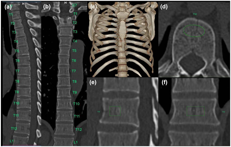
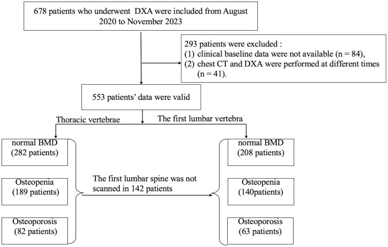
Methods: A total of 553 participants (353 men and 200 women) who underwent chest CT and DXA within 1 day were included. The attenuation values (HU) of the T7-12 vertebrae and L1 vertebra were obtained by AI. The effects of the clinical baseline data and attenuation values among the normal, osteopenia, and osteoporosis groups were compared. The correlation between attenuation and bone mineral density (BMD) values was analyzed, and the diagnostic performance of thoracic and first lumbar vertebrae attenuation values for diagnosing osteopenia or osteoporosis was further explored.
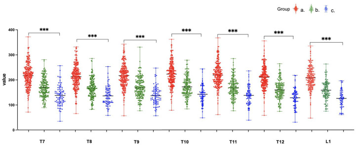
Results: The CT attenuation values of T7-12 and L1 vertebrae showed positive correlation with T-score (R = 0.58-0.61, p < 0.01). T12 attenuation >184.8 HU was 84.1% sensitive and 70.6% specific for distinguishing normal BMD, while T12 attenuation <146.2 HU was 61.4% specific and 75.6% sensitive for distinguishing osteoporosis from osteopenia. There were no significant differences between the T10-12 and L1 groups in distinguishing the normal, osteopenia, and osteoporosis groups. Moreover, the diagnostic efficacy among the T10, T11, T12, and L1 vertebral bodies was not statistically significantly different among the three groups.
Conclusion: Opportunistic screening is a valid method for predicting osteopenia or osteoporosis. As a rapid and effective tool, T10-12 vertebral attenuation measures can be incorporated to predict osteoporosis and identify patients who may benefit from further investigations using DXA based on routine clinical chest CT examinations.
期刊介绍:
Therapeutic Advances in Musculoskeletal Disease delivers the highest quality peer-reviewed articles, reviews, and scholarly comment on pioneering efforts and innovative studies across all areas of musculoskeletal disease.
 求助内容:
求助内容: 应助结果提醒方式:


