通过枕外突形态判断性别:印度西北部的CT扫描研究
IF 1
Q4 RADIOLOGY, NUCLEAR MEDICINE & MEDICAL IMAGING
引用次数: 0
摘要
目的:本研究旨在利用计算机断层扫描(CT)评估西北印度人群中枕外隆突(EOP)形态学在性别估计中的类型、流行程度和法医应用。材料与方法对年龄在18 ~ 75岁的331例成人(男性167例,女性164例)的ct扫描数据进行分析。EOP形态分为三种不同的类型:1型、2型和3型。形态学测量-包括EOP总长度(TEOP)和EOP顶点角度(AAEOP) -使用Radiant DICOM Viewer获得。采用统计分析,包括判别函数分析,评估EOP特征对性别估计的预测准确性。结果EOP形态存在明显的性别二态性。1型多见于女性(33.8%),3型多见于男性(38.3%)。男性个体表现出更大的EOP厚度和更明显的颅标志,如上、下颈线和枕外嵴。判别函数分析的总体性别估计准确率为89.4%,在年龄较大的年龄组中分类率较高。其中,TEOP和AAEOP是最可靠的预测因子,分类准确率分别为81.9%和76.1%。结论eop形态学表现出明显的性别二态性,可作为西北印第安人性别估计的可靠指标。高分类准确性,特别是在老年群体中,支持将基于eop的评估整合到法医协议中。本研究强调了人口特异性数据的重要性,并鼓励在代表性不足的地区进一步研究颅骨特征,以提高法医鉴定方法。本文章由计算机程序翻译,如有差异,请以英文原文为准。
Sex Estimation Through External Occipital Protuberance Morphology: A CT Scan Study in Northwest India
Objectives
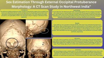
This study aims to evaluate the types, prevalence, and forensic utility of external occipital protuberance (EOP) morphology for sex estimation in a Northwest Indian population using computed tomography (CT) imaging.
Materials and Methods
CT scan data of 331 adult individuals (167 males, 164 females), aged 18 to 75 years, were analyzed. EOP morphology was classified into three distinct types: Type 1, Type 2, and Type 3. Morphometric measurements—including total EOP length (TEOP) and angle at apex of EOP (AAEOP)—were obtained using the Radiant DICOM Viewer. Statistical analysis, including discriminant function analysis, was employed to assess the predictive accuracy of EOP features for sex estimation.
Results
The analysis revealed significant sexual dimorphism in EOP morphology. Type 1 was more frequent in females (33.8%), while Type 3 was more common in males (38.3%). Male individuals showed greater EOP thickness and more pronounced cranial landmarks such as the superior and inferior nuchal lines and the external occipital crest. Discriminant function analysis yielded an overall sex estimation accuracy of 89.4%, with higher classification rates in older age groups. Among the variables, TEOP and AAEOP were the most reliable predictors, achieving classification accuracies of 81.9% and 76.1%, respectively.
Conclusion
EOP morphology demonstrates clear sexual dimorphism and proves to be a reliable indicator for sex estimation in the studied Northwest Indian population. The high classification accuracy, particularly in older age groups, supports the integration of EOP-based assessment into forensic protocols. This study highlights the importance of population-specific data and encourages further research on cranial features in underrepresented regions to enhance forensic identification methods.
求助全文
通过发布文献求助,成功后即可免费获取论文全文。
去求助
来源期刊
Forensic Imaging
RADIOLOGY, NUCLEAR MEDICINE & MEDICAL IMAGING-
CiteScore
2.20
自引率
27.30%
发文量
39
 求助内容:
求助内容: 应助结果提醒方式:


