{"title":"基于ct的放射组学模型解码胰腺导管腺癌的纤维化内容和分子差异:一项多机构研究。","authors":"Fangqing Wang, Yang Sun, Jianwei Xu, Yufan Chen, Hui Zhang, Guotao Yin, Dexin Yu","doi":"10.1186/s13244-025-02036-z","DOIUrl":null,"url":null,"abstract":"<p><strong>Objectives: </strong>To develop a CT radiomics model for predicting fibrosis grade in pancreatic ductal adenocarcinoma (PDAC) and to investigate the underlying prognosis value and biological basis.</p><p><strong>Methods: </strong>Patients with resected PDAC were retrospectively included from three institutions. Evaluating tumor fibrosis content using fibrotic pixels proportion through Masson staining of postoperative pathological sections. Radiomics features from preoperative contrast-enhanced CT (CECT) were extracted and used to develop models in the training cohort. The diagnosis performance was further validated in the two test cohorts. The outcome cohort, including patients with advanced PDAC undergoing neoadjuvant chemotherapy, was used to evaluate the predictive value of the model for overall survival (OS) and disease-free survival (DFS), which were investigated using the Kaplan-Meier method and log-rank test. RNA sequencing data from a prospective biological basis cohort were conducted to explore the biological processes underlying the radiomics model.</p><p><strong>Results: </strong>Among 215 patients (median age 60.89 years, 142 men) used for radiomics modeling, 132 (61.40%) were confirmed as high fibrosis content. The combined phase (CP) radiomics model, which included all CECT radiomics features, showed the best performance for predicting fibrosis grade, with AUCs of 0.831, 0.785, and 0.746 in training, internal test, and external test cohorts. OS (p = 0.011) and DFS (p = 0.022) can be categorized using the CP radiomics model in the outcome cohort. RNA-seq indicated that different CP models were associated with fibrotic production and remodeling processes.</p><p><strong>Conclusion: </strong>The CP radiomics model showed the best performance in predicting fibrosis grades in PDAC.</p><p><strong>Critical relevance statement: </strong>Fibrosis grading is of prognostic and neoadjuvant chemotherapy efficacy evaluation significance, and the CT-based combined phase radiomics model established in our study will facilitate risk stratification and selection of personalized treatment strategies for patients. Furthermore, underlying biological processes demonstrated in the radiomics model will offer valuable insights into their interpretability and clinical translation.</p><p><strong>Key points: </strong>Fibrosis grading is of prognostic significance in pancreatic ductal adenocarcinoma (PDAC), but lacks a reliable preoperative assessment. The CT-based combined phase (CP) radiomics model predicts fibrosis grading effectively in PDAC. The CP radiomics model demonstrated prognostic and neoadjuvant chemotherapy efficacy evaluation value and underlying biological processes, which related fibrotic production and remodeling processes.</p>","PeriodicalId":13639,"journal":{"name":"Insights into Imaging","volume":"16 1","pages":"190"},"PeriodicalIF":4.5000,"publicationDate":"2025-09-12","publicationTypes":"Journal Article","fieldsOfStudy":null,"isOpenAccess":false,"openAccessPdf":"https://www.ncbi.nlm.nih.gov/pmc/articles/PMC12431998/pdf/","citationCount":"0","resultStr":"{\"title\":\"CT-based radiomics models decode fibrosis content and molecular differences in pancreatic ductal adenocarcinoma: a multi-institutional study.\",\"authors\":\"Fangqing Wang, Yang Sun, Jianwei Xu, Yufan Chen, Hui Zhang, Guotao Yin, Dexin Yu\",\"doi\":\"10.1186/s13244-025-02036-z\",\"DOIUrl\":null,\"url\":null,\"abstract\":\"<p><strong>Objectives: </strong>To develop a CT radiomics model for predicting fibrosis grade in pancreatic ductal adenocarcinoma (PDAC) and to investigate the underlying prognosis value and biological basis.</p><p><strong>Methods: </strong>Patients with resected PDAC were retrospectively included from three institutions. Evaluating tumor fibrosis content using fibrotic pixels proportion through Masson staining of postoperative pathological sections. Radiomics features from preoperative contrast-enhanced CT (CECT) were extracted and used to develop models in the training cohort. The diagnosis performance was further validated in the two test cohorts. The outcome cohort, including patients with advanced PDAC undergoing neoadjuvant chemotherapy, was used to evaluate the predictive value of the model for overall survival (OS) and disease-free survival (DFS), which were investigated using the Kaplan-Meier method and log-rank test. RNA sequencing data from a prospective biological basis cohort were conducted to explore the biological processes underlying the radiomics model.</p><p><strong>Results: </strong>Among 215 patients (median age 60.89 years, 142 men) used for radiomics modeling, 132 (61.40%) were confirmed as high fibrosis content. The combined phase (CP) radiomics model, which included all CECT radiomics features, showed the best performance for predicting fibrosis grade, with AUCs of 0.831, 0.785, and 0.746 in training, internal test, and external test cohorts. OS (p = 0.011) and DFS (p = 0.022) can be categorized using the CP radiomics model in the outcome cohort. RNA-seq indicated that different CP models were associated with fibrotic production and remodeling processes.</p><p><strong>Conclusion: </strong>The CP radiomics model showed the best performance in predicting fibrosis grades in PDAC.</p><p><strong>Critical relevance statement: </strong>Fibrosis grading is of prognostic and neoadjuvant chemotherapy efficacy evaluation significance, and the CT-based combined phase radiomics model established in our study will facilitate risk stratification and selection of personalized treatment strategies for patients. Furthermore, underlying biological processes demonstrated in the radiomics model will offer valuable insights into their interpretability and clinical translation.</p><p><strong>Key points: </strong>Fibrosis grading is of prognostic significance in pancreatic ductal adenocarcinoma (PDAC), but lacks a reliable preoperative assessment. The CT-based combined phase (CP) radiomics model predicts fibrosis grading effectively in PDAC. The CP radiomics model demonstrated prognostic and neoadjuvant chemotherapy efficacy evaluation value and underlying biological processes, which related fibrotic production and remodeling processes.</p>\",\"PeriodicalId\":13639,\"journal\":{\"name\":\"Insights into Imaging\",\"volume\":\"16 1\",\"pages\":\"190\"},\"PeriodicalIF\":4.5000,\"publicationDate\":\"2025-09-12\",\"publicationTypes\":\"Journal Article\",\"fieldsOfStudy\":null,\"isOpenAccess\":false,\"openAccessPdf\":\"https://www.ncbi.nlm.nih.gov/pmc/articles/PMC12431998/pdf/\",\"citationCount\":\"0\",\"resultStr\":null,\"platform\":\"Semanticscholar\",\"paperid\":null,\"PeriodicalName\":\"Insights into Imaging\",\"FirstCategoryId\":\"3\",\"ListUrlMain\":\"https://doi.org/10.1186/s13244-025-02036-z\",\"RegionNum\":2,\"RegionCategory\":\"医学\",\"ArticlePicture\":[],\"TitleCN\":null,\"AbstractTextCN\":null,\"PMCID\":null,\"EPubDate\":\"\",\"PubModel\":\"\",\"JCR\":\"Q1\",\"JCRName\":\"RADIOLOGY, NUCLEAR MEDICINE & MEDICAL IMAGING\",\"Score\":null,\"Total\":0}","platform":"Semanticscholar","paperid":null,"PeriodicalName":"Insights into Imaging","FirstCategoryId":"3","ListUrlMain":"https://doi.org/10.1186/s13244-025-02036-z","RegionNum":2,"RegionCategory":"医学","ArticlePicture":[],"TitleCN":null,"AbstractTextCN":null,"PMCID":null,"EPubDate":"","PubModel":"","JCR":"Q1","JCRName":"RADIOLOGY, NUCLEAR MEDICINE & MEDICAL IMAGING","Score":null,"Total":0}
CT-based radiomics models decode fibrosis content and molecular differences in pancreatic ductal adenocarcinoma: a multi-institutional study.
Objectives: To develop a CT radiomics model for predicting fibrosis grade in pancreatic ductal adenocarcinoma (PDAC) and to investigate the underlying prognosis value and biological basis.
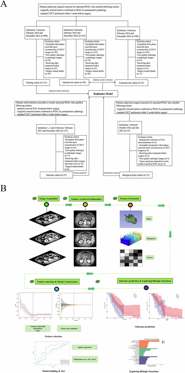
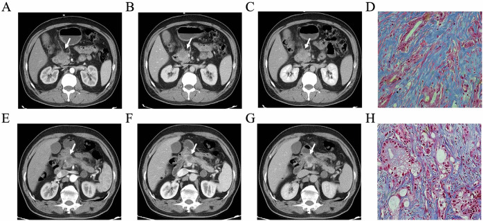
Methods: Patients with resected PDAC were retrospectively included from three institutions. Evaluating tumor fibrosis content using fibrotic pixels proportion through Masson staining of postoperative pathological sections. Radiomics features from preoperative contrast-enhanced CT (CECT) were extracted and used to develop models in the training cohort. The diagnosis performance was further validated in the two test cohorts. The outcome cohort, including patients with advanced PDAC undergoing neoadjuvant chemotherapy, was used to evaluate the predictive value of the model for overall survival (OS) and disease-free survival (DFS), which were investigated using the Kaplan-Meier method and log-rank test. RNA sequencing data from a prospective biological basis cohort were conducted to explore the biological processes underlying the radiomics model.
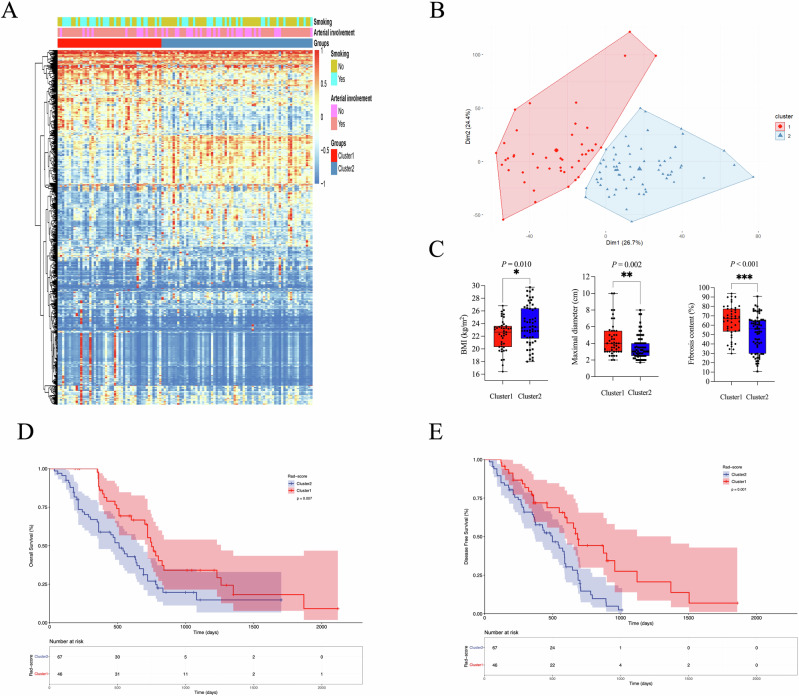
Results: Among 215 patients (median age 60.89 years, 142 men) used for radiomics modeling, 132 (61.40%) were confirmed as high fibrosis content. The combined phase (CP) radiomics model, which included all CECT radiomics features, showed the best performance for predicting fibrosis grade, with AUCs of 0.831, 0.785, and 0.746 in training, internal test, and external test cohorts. OS (p = 0.011) and DFS (p = 0.022) can be categorized using the CP radiomics model in the outcome cohort. RNA-seq indicated that different CP models were associated with fibrotic production and remodeling processes.
Conclusion: The CP radiomics model showed the best performance in predicting fibrosis grades in PDAC.
Critical relevance statement: Fibrosis grading is of prognostic and neoadjuvant chemotherapy efficacy evaluation significance, and the CT-based combined phase radiomics model established in our study will facilitate risk stratification and selection of personalized treatment strategies for patients. Furthermore, underlying biological processes demonstrated in the radiomics model will offer valuable insights into their interpretability and clinical translation.
Key points: Fibrosis grading is of prognostic significance in pancreatic ductal adenocarcinoma (PDAC), but lacks a reliable preoperative assessment. The CT-based combined phase (CP) radiomics model predicts fibrosis grading effectively in PDAC. The CP radiomics model demonstrated prognostic and neoadjuvant chemotherapy efficacy evaluation value and underlying biological processes, which related fibrotic production and remodeling processes.
期刊介绍:
Insights into Imaging (I³) is a peer-reviewed open access journal published under the brand SpringerOpen. All content published in the journal is freely available online to anyone, anywhere!
I³ continuously updates scientific knowledge and progress in best-practice standards in radiology through the publication of original articles and state-of-the-art reviews and opinions, along with recommendations and statements from the leading radiological societies in Europe.
Founded by the European Society of Radiology (ESR), I³ creates a platform for educational material, guidelines and recommendations, and a forum for topics of controversy.
A balanced combination of review articles, original papers, short communications from European radiological congresses and information on society matters makes I³ an indispensable source for current information in this field.
I³ is owned by the ESR, however authors retain copyright to their article according to the Creative Commons Attribution License (see Copyright and License Agreement). All articles can be read, redistributed and reused for free, as long as the author of the original work is cited properly.
The open access fees (article-processing charges) for this journal are kindly sponsored by ESR for all Members.
The journal went open access in 2012, which means that all articles published since then are freely available online.

 求助内容:
求助内容: 应助结果提醒方式:


