Ziyun Liang , Xiaoqing Guo , Wentian Xu , Yasin Ibrahim , Natalie Voets , Pieter M. Pretorius , Alzheimer’s Disease Neuroimaging Initiative, J. Alison Noble , Konstantinos Kamnitsas
{"title":"IterMask3D:基于测试时间迭代掩模细化的三维脑MRI无监督异常检测与分割","authors":"Ziyun Liang , Xiaoqing Guo , Wentian Xu , Yasin Ibrahim , Natalie Voets , Pieter M. Pretorius , Alzheimer’s Disease Neuroimaging Initiative, J. Alison Noble , Konstantinos Kamnitsas","doi":"10.1016/j.media.2025.103763","DOIUrl":null,"url":null,"abstract":"<div><div>Unsupervised anomaly detection and segmentation methods train a model to learn the training distribution as ‘normal’. In the testing phase, they identify patterns that deviate from this normal distribution as ‘anomalies’. To learn the ‘normal’ distribution, prevailing methods corrupt the images and train a model to reconstruct them. During testing, the model attempts to reconstruct corrupted inputs based on the learned ‘normal’ distribution. Deviations from this distribution lead to high reconstruction errors, which indicate potential anomalies. However, corrupting an input image inevitably causes information loss even in normal regions, leading to suboptimal reconstruction and an increased risk of false positives. To alleviate this, we propose <span><math><mi>IterMask3D</mi></math></span>, an iterative spatial mask-refining strategy designed for 3D brain MRI. We iteratively spatially mask areas of the image as corruption and reconstruct them, then shrink the mask based on reconstruction error. This process iteratively unmasks ‘normal’ areas to the model, whose information further guides reconstruction of ‘normal’ patterns under the mask to be reconstructed accurately, reducing false positives. In addition, to achieve better reconstruction performance, we also propose using high-frequency image content as additional structural information to guide the reconstruction of the masked area. Extensive experiments on the detection of both synthetic and real-world imaging artifacts, as well as segmentation of various pathological lesions across multiple MRI sequences, consistently demonstrate the effectiveness of our proposed method. Code is available at <span><span>https://github.com/ZiyunLiang/IterMask3D</span><svg><path></path></svg></span>.</div></div>","PeriodicalId":18328,"journal":{"name":"Medical image analysis","volume":"107 ","pages":"Article 103763"},"PeriodicalIF":11.8000,"publicationDate":"2025-08-28","publicationTypes":"Journal Article","fieldsOfStudy":null,"isOpenAccess":false,"openAccessPdf":"","citationCount":"0","resultStr":"{\"title\":\"IterMask3D: Unsupervised anomaly detection and segmentation with test-time iterative mask refinement in 3D brain MRI\",\"authors\":\"Ziyun Liang , Xiaoqing Guo , Wentian Xu , Yasin Ibrahim , Natalie Voets , Pieter M. Pretorius , Alzheimer’s Disease Neuroimaging Initiative, J. Alison Noble , Konstantinos Kamnitsas\",\"doi\":\"10.1016/j.media.2025.103763\",\"DOIUrl\":null,\"url\":null,\"abstract\":\"<div><div>Unsupervised anomaly detection and segmentation methods train a model to learn the training distribution as ‘normal’. In the testing phase, they identify patterns that deviate from this normal distribution as ‘anomalies’. To learn the ‘normal’ distribution, prevailing methods corrupt the images and train a model to reconstruct them. During testing, the model attempts to reconstruct corrupted inputs based on the learned ‘normal’ distribution. Deviations from this distribution lead to high reconstruction errors, which indicate potential anomalies. However, corrupting an input image inevitably causes information loss even in normal regions, leading to suboptimal reconstruction and an increased risk of false positives. To alleviate this, we propose <span><math><mi>IterMask3D</mi></math></span>, an iterative spatial mask-refining strategy designed for 3D brain MRI. We iteratively spatially mask areas of the image as corruption and reconstruct them, then shrink the mask based on reconstruction error. This process iteratively unmasks ‘normal’ areas to the model, whose information further guides reconstruction of ‘normal’ patterns under the mask to be reconstructed accurately, reducing false positives. In addition, to achieve better reconstruction performance, we also propose using high-frequency image content as additional structural information to guide the reconstruction of the masked area. Extensive experiments on the detection of both synthetic and real-world imaging artifacts, as well as segmentation of various pathological lesions across multiple MRI sequences, consistently demonstrate the effectiveness of our proposed method. Code is available at <span><span>https://github.com/ZiyunLiang/IterMask3D</span><svg><path></path></svg></span>.</div></div>\",\"PeriodicalId\":18328,\"journal\":{\"name\":\"Medical image analysis\",\"volume\":\"107 \",\"pages\":\"Article 103763\"},\"PeriodicalIF\":11.8000,\"publicationDate\":\"2025-08-28\",\"publicationTypes\":\"Journal Article\",\"fieldsOfStudy\":null,\"isOpenAccess\":false,\"openAccessPdf\":\"\",\"citationCount\":\"0\",\"resultStr\":null,\"platform\":\"Semanticscholar\",\"paperid\":null,\"PeriodicalName\":\"Medical image analysis\",\"FirstCategoryId\":\"5\",\"ListUrlMain\":\"https://www.sciencedirect.com/science/article/pii/S1361841525003093\",\"RegionNum\":1,\"RegionCategory\":\"医学\",\"ArticlePicture\":[],\"TitleCN\":null,\"AbstractTextCN\":null,\"PMCID\":null,\"EPubDate\":\"\",\"PubModel\":\"\",\"JCR\":\"Q1\",\"JCRName\":\"COMPUTER SCIENCE, ARTIFICIAL INTELLIGENCE\",\"Score\":null,\"Total\":0}","platform":"Semanticscholar","paperid":null,"PeriodicalName":"Medical image analysis","FirstCategoryId":"5","ListUrlMain":"https://www.sciencedirect.com/science/article/pii/S1361841525003093","RegionNum":1,"RegionCategory":"医学","ArticlePicture":[],"TitleCN":null,"AbstractTextCN":null,"PMCID":null,"EPubDate":"","PubModel":"","JCR":"Q1","JCRName":"COMPUTER SCIENCE, ARTIFICIAL INTELLIGENCE","Score":null,"Total":0}
IterMask3D: Unsupervised anomaly detection and segmentation with test-time iterative mask refinement in 3D brain MRI
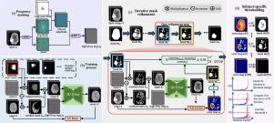
Unsupervised anomaly detection and segmentation methods train a model to learn the training distribution as ‘normal’. In the testing phase, they identify patterns that deviate from this normal distribution as ‘anomalies’. To learn the ‘normal’ distribution, prevailing methods corrupt the images and train a model to reconstruct them. During testing, the model attempts to reconstruct corrupted inputs based on the learned ‘normal’ distribution. Deviations from this distribution lead to high reconstruction errors, which indicate potential anomalies. However, corrupting an input image inevitably causes information loss even in normal regions, leading to suboptimal reconstruction and an increased risk of false positives. To alleviate this, we propose , an iterative spatial mask-refining strategy designed for 3D brain MRI. We iteratively spatially mask areas of the image as corruption and reconstruct them, then shrink the mask based on reconstruction error. This process iteratively unmasks ‘normal’ areas to the model, whose information further guides reconstruction of ‘normal’ patterns under the mask to be reconstructed accurately, reducing false positives. In addition, to achieve better reconstruction performance, we also propose using high-frequency image content as additional structural information to guide the reconstruction of the masked area. Extensive experiments on the detection of both synthetic and real-world imaging artifacts, as well as segmentation of various pathological lesions across multiple MRI sequences, consistently demonstrate the effectiveness of our proposed method. Code is available at https://github.com/ZiyunLiang/IterMask3D.
期刊介绍:
Medical Image Analysis serves as a platform for sharing new research findings in the realm of medical and biological image analysis, with a focus on applications of computer vision, virtual reality, and robotics to biomedical imaging challenges. The journal prioritizes the publication of high-quality, original papers contributing to the fundamental science of processing, analyzing, and utilizing medical and biological images. It welcomes approaches utilizing biomedical image datasets across all spatial scales, from molecular/cellular imaging to tissue/organ imaging.