Ofeye 3.0软件程序的开发和多中心验证,用于通过胸部/腹部CT图像自动进行全方位椎体骨折检测
IF 5.9
1区 医学
Q1 ORTHOPEDICS
引用次数: 0
摘要
背景:当适应症不是脊柱疾病时,胸腹CT上脆性椎体骨折(VF)的缺失报告很常见。这意味着错过了提醒患者采取预防措施以改善骨骼健康和防止进一步严重骨折的机会。在这项研究中,我们的目标是开发一个软件程序来自动检测VF与现有的胸部/腹部CT扫描,并验证其检测性能。方法建立CT轴向图像自动矢状中央板重建(CSR)方法。作为参考VF读数,确定VF包括椎体高度损失20%的VF和椎体高度损失最小的终板骨折VF。VFs也可与骨关节炎楔入性和短椎骨内炎区分。将侧位x线片VF检测模型的先验知识转移到新的“Ofeye 3.0”模型中,该模型针对CT图像的VF检测进行了优化。从9个中心获得训练CT图像,无VF 3313例,有VF 835例。为了进行外部验证,CT图像来自5个中心,共732例无VF和224例有VF。结果自动CSR方法在显示终板及邻近结构的结构变化方面具有优势。对于胸/腹CT扫描中VF的检测,逐例计数并与参考读数比较,Ofeye 3.0的平均表现为准确率0.967,灵敏度0.906,特异性0.986。大多数假阴性或假阳性病例是轻微或轻微的VF,或有图像伪影,或VF靠近CSR图像的外围。尽管对软件检测全面VF的要求具有挑战性,但我们的结果与其他已发表的自动VF检测模型相比具有优势。我们开发了一个软件程序,用于自动检测胸部和/或腹部CT图像数据的全方位VF,并进行了多中心外部验证研究。实验证明该软件具有较高的VF检测精度。通过提醒患者可能与骨质疏松症相关的VFs,从而使患者采取措施防止进一步骨折,将该软件集成到放射实践中将改善患者的治疗效果并降低医疗成本。本文章由计算机程序翻译,如有差异,请以英文原文为准。
Development and multi-center validation of a software program, Ofeye 3.0, for automated all-inclusive vertebral fracture detection with chest/abdominal CT images
Background
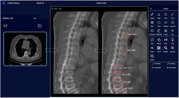
Missing report for fragility vertebral fracture (VF) on chest/abdominal CT is common when the indication is not spine disorders. This represents a missed opportunity to alert patients to take preventive measures to improve bone health and prevent further severe fractures. In this study, we aim to develop a software program for automated detection of VF with existing chest/abdominal CT scans and validate its detection performance.
Methods
An automated sagittal Central Slab reconstruction (CSR) method for CT axial images was developed. For reference VF reading, VFs inclusive of those of with <20 % vertebral height loss and those of endplate fracture with minimal vertebral height loss were identified. VFs were also differentiated from osteoarthritic wedging and endplatitis short vertebrae. Prior knowledge of VF detection models for lateral radiograph were transferred to a new ‘Ofeye 3.0’ model optimized for VF detection on CT image. Training CT images were obtained from nine centers, totaling 3313 cases without VF and 835 cases with VF. For external validation, CT images were from five centers totaling 732 cases without VF and 224 cases with VF.
Results
The automated CSR method showed advantages in demonstrating structural changes of the endplate and adjacent structures. For detecting VF in chest/abdominal CT scans, counting case-by-case and compared with the reference reading, the average performance of Ofeye 3.0 was accuracy 0.967, sensitivity 0.906, and specificity 0.986. Most of false negative or false positive cases were minimal or mild VF, or with image artifacts, or with VF close to the peripheral of CSR images.
Conclusions
Despite the challenging requirements for the software to detect all-inclusive VF, our results compare favorably with other published automated VF detection models.
The translational potential of this article
We developed a software program for automated all-inclusive VF detection on chest and/or abdominal CT image data and conducted a multi-center external validation study. This software is proved to have high VF detection precision. By alerting patients of the VFs likely related to osteoporosis and in turn the patients taking measures to prevent further fracture, the integration of this software into radiological practice will improve patient outcomes and reduce healthcare costs.
求助全文
通过发布文献求助,成功后即可免费获取论文全文。
去求助
来源期刊
Journal of Orthopaedic Translation
Medicine-Orthopedics and Sports Medicine
CiteScore
11.80
自引率
13.60%
发文量
91
审稿时长
29 days
期刊介绍:
The Journal of Orthopaedic Translation (JOT) is the official peer-reviewed, open access journal of the Chinese Speaking Orthopaedic Society (CSOS) and the International Chinese Musculoskeletal Research Society (ICMRS). It is published quarterly, in January, April, July and October, by Elsevier.
 求助内容:
求助内容: 应助结果提醒方式:


