Eve Lennie, Steven Sourbron, Nigel Hoggard, Thomas Jenkins, Charalampos Tsoumpas
{"title":"PET/MRI定量脊髓FDG。","authors":"Eve Lennie, Steven Sourbron, Nigel Hoggard, Thomas Jenkins, Charalampos Tsoumpas","doi":"10.3389/fnume.2025.1646662","DOIUrl":null,"url":null,"abstract":"<p><strong>Background: </strong>In this study, we investigate the impact of MR-derived attenuation maps and limited detector resolution on the quantification of positron emission tomography (PET) activity uptake in the spinal cord during PET/MRI. This was performed by simulating [ <math><msup><mi></mi> <mrow><mn>18</mn></mrow> </msup> </math> F]FDG PET data in the neck and thorax and then modifying the attenuation map to remove bone features. We then compared Ordered Subset Expectation Maximisation-reconstructed images to those with full attenuation correction. This simulation was performed at two detector resolutions of 2.1 and 4.4 mm. Acquisitions from a clinical study were then used to assess the ability of point spread function (PSF) modelling and time-of-flight (TOF) corrections, as implemented on the SIGNA PET/MR scanner (GE HealthCare), to correct for these quantification errors. For comparison, mean uptake was measured in regions of interest at each vertebral position along the spinal cord.</p><p><strong>Results: </strong>Simulation results showed a decreasing pattern of uptake from the cervical to the thoracic spinal cord. When bone was not included in attenuation correction, the mean uptake decreased by 3%-10.4%. This difference in measured uptake was 6.4%-23.9% in images simulated at a detector resolution representative of a clinical PET/MRI scanner. At a detector resolution of 4.4 mm, a 32.2% decrease in uptake was measured compared to the 2.1 mm simulation. In patient data, introducing vertebral bone to the attenuation correction pseudo-CT led to a 1.8%-18.3% difference in <math> <msub><mrow><mi>SUV</mi></mrow> <mrow><mrow><mi>mean</mi></mrow> </mrow> </msub> </math> in the spinal cord. Applying PSF modelling did not lead to any statistically significant changes. TOF correction reduces the difference in <math> <msub><mrow><mi>SUV</mi></mrow> <mrow><mrow><mi>mean</mi></mrow> </mrow> </msub> </math> between data attenuation corrected with and without vertebral bone to 4.3%-7%. TOF Q.Clear images with beta = 100 showed the smallest difference between attenuation correction approaches at 0.6%-5.2%.</p><p><strong>Conclusion: </strong>Ignoring bone during image reconstruction in PET/MRI reduces the activity measured during quantification of the spinal cord; however, the partial volume effect has a greater impact on reducing measured uptake in lower-resolution data. While time-of-flight correction goes somewhat resolves these quantification errors, further research is needed into partial volume correction.</p>","PeriodicalId":73095,"journal":{"name":"Frontiers in nuclear medicine (Lausanne, Switzerland)","volume":"5 ","pages":"1646662"},"PeriodicalIF":1.4000,"publicationDate":"2025-08-26","publicationTypes":"Journal Article","fieldsOfStudy":null,"isOpenAccess":false,"openAccessPdf":"https://www.ncbi.nlm.nih.gov/pmc/articles/PMC12417479/pdf/","citationCount":"0","resultStr":"{\"title\":\"Quantification of FDG in the spinal cord using PET/MRI.\",\"authors\":\"Eve Lennie, Steven Sourbron, Nigel Hoggard, Thomas Jenkins, Charalampos Tsoumpas\",\"doi\":\"10.3389/fnume.2025.1646662\",\"DOIUrl\":null,\"url\":null,\"abstract\":\"<p><strong>Background: </strong>In this study, we investigate the impact of MR-derived attenuation maps and limited detector resolution on the quantification of positron emission tomography (PET) activity uptake in the spinal cord during PET/MRI. This was performed by simulating [ <math><msup><mi></mi> <mrow><mn>18</mn></mrow> </msup> </math> F]FDG PET data in the neck and thorax and then modifying the attenuation map to remove bone features. We then compared Ordered Subset Expectation Maximisation-reconstructed images to those with full attenuation correction. This simulation was performed at two detector resolutions of 2.1 and 4.4 mm. Acquisitions from a clinical study were then used to assess the ability of point spread function (PSF) modelling and time-of-flight (TOF) corrections, as implemented on the SIGNA PET/MR scanner (GE HealthCare), to correct for these quantification errors. For comparison, mean uptake was measured in regions of interest at each vertebral position along the spinal cord.</p><p><strong>Results: </strong>Simulation results showed a decreasing pattern of uptake from the cervical to the thoracic spinal cord. When bone was not included in attenuation correction, the mean uptake decreased by 3%-10.4%. This difference in measured uptake was 6.4%-23.9% in images simulated at a detector resolution representative of a clinical PET/MRI scanner. At a detector resolution of 4.4 mm, a 32.2% decrease in uptake was measured compared to the 2.1 mm simulation. In patient data, introducing vertebral bone to the attenuation correction pseudo-CT led to a 1.8%-18.3% difference in <math> <msub><mrow><mi>SUV</mi></mrow> <mrow><mrow><mi>mean</mi></mrow> </mrow> </msub> </math> in the spinal cord. Applying PSF modelling did not lead to any statistically significant changes. TOF correction reduces the difference in <math> <msub><mrow><mi>SUV</mi></mrow> <mrow><mrow><mi>mean</mi></mrow> </mrow> </msub> </math> between data attenuation corrected with and without vertebral bone to 4.3%-7%. TOF Q.Clear images with beta = 100 showed the smallest difference between attenuation correction approaches at 0.6%-5.2%.</p><p><strong>Conclusion: </strong>Ignoring bone during image reconstruction in PET/MRI reduces the activity measured during quantification of the spinal cord; however, the partial volume effect has a greater impact on reducing measured uptake in lower-resolution data. While time-of-flight correction goes somewhat resolves these quantification errors, further research is needed into partial volume correction.</p>\",\"PeriodicalId\":73095,\"journal\":{\"name\":\"Frontiers in nuclear medicine (Lausanne, Switzerland)\",\"volume\":\"5 \",\"pages\":\"1646662\"},\"PeriodicalIF\":1.4000,\"publicationDate\":\"2025-08-26\",\"publicationTypes\":\"Journal Article\",\"fieldsOfStudy\":null,\"isOpenAccess\":false,\"openAccessPdf\":\"https://www.ncbi.nlm.nih.gov/pmc/articles/PMC12417479/pdf/\",\"citationCount\":\"0\",\"resultStr\":null,\"platform\":\"Semanticscholar\",\"paperid\":null,\"PeriodicalName\":\"Frontiers in nuclear medicine (Lausanne, Switzerland)\",\"FirstCategoryId\":\"1085\",\"ListUrlMain\":\"https://doi.org/10.3389/fnume.2025.1646662\",\"RegionNum\":0,\"RegionCategory\":null,\"ArticlePicture\":[],\"TitleCN\":null,\"AbstractTextCN\":null,\"PMCID\":null,\"EPubDate\":\"2025/1/1 0:00:00\",\"PubModel\":\"eCollection\",\"JCR\":\"\",\"JCRName\":\"\",\"Score\":null,\"Total\":0}","platform":"Semanticscholar","paperid":null,"PeriodicalName":"Frontiers in nuclear medicine (Lausanne, Switzerland)","FirstCategoryId":"1085","ListUrlMain":"https://doi.org/10.3389/fnume.2025.1646662","RegionNum":0,"RegionCategory":null,"ArticlePicture":[],"TitleCN":null,"AbstractTextCN":null,"PMCID":null,"EPubDate":"2025/1/1 0:00:00","PubModel":"eCollection","JCR":"","JCRName":"","Score":null,"Total":0}
Quantification of FDG in the spinal cord using PET/MRI.
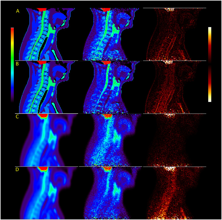
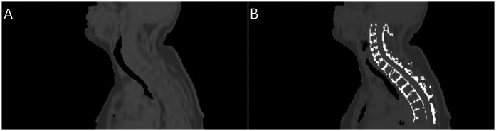
Background: In this study, we investigate the impact of MR-derived attenuation maps and limited detector resolution on the quantification of positron emission tomography (PET) activity uptake in the spinal cord during PET/MRI. This was performed by simulating [ F]FDG PET data in the neck and thorax and then modifying the attenuation map to remove bone features. We then compared Ordered Subset Expectation Maximisation-reconstructed images to those with full attenuation correction. This simulation was performed at two detector resolutions of 2.1 and 4.4 mm. Acquisitions from a clinical study were then used to assess the ability of point spread function (PSF) modelling and time-of-flight (TOF) corrections, as implemented on the SIGNA PET/MR scanner (GE HealthCare), to correct for these quantification errors. For comparison, mean uptake was measured in regions of interest at each vertebral position along the spinal cord.
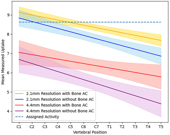
Results: Simulation results showed a decreasing pattern of uptake from the cervical to the thoracic spinal cord. When bone was not included in attenuation correction, the mean uptake decreased by 3%-10.4%. This difference in measured uptake was 6.4%-23.9% in images simulated at a detector resolution representative of a clinical PET/MRI scanner. At a detector resolution of 4.4 mm, a 32.2% decrease in uptake was measured compared to the 2.1 mm simulation. In patient data, introducing vertebral bone to the attenuation correction pseudo-CT led to a 1.8%-18.3% difference in in the spinal cord. Applying PSF modelling did not lead to any statistically significant changes. TOF correction reduces the difference in between data attenuation corrected with and without vertebral bone to 4.3%-7%. TOF Q.Clear images with beta = 100 showed the smallest difference between attenuation correction approaches at 0.6%-5.2%.
Conclusion: Ignoring bone during image reconstruction in PET/MRI reduces the activity measured during quantification of the spinal cord; however, the partial volume effect has a greater impact on reducing measured uptake in lower-resolution data. While time-of-flight correction goes somewhat resolves these quantification errors, further research is needed into partial volume correction.

 求助内容:
求助内容: 应助结果提醒方式:


