Robin Litten, James Pate, Austin Hughes, Jordan Dunson, Chadwick Smith, Jeremy Bruce
{"title":"前交叉韧带修复后内翻性独眼病变1例。","authors":"Robin Litten, James Pate, Austin Hughes, Jordan Dunson, Chadwick Smith, Jeremy Bruce","doi":"10.13107/jocr.2025.v15.i09.6050","DOIUrl":null,"url":null,"abstract":"<p><strong>Introduction: </strong>Cyclops lesions are a well-described complication following reconstruction of the anterior cruciate ligament (ACL). These lesions are fibrous nodules that most commonly form anterolateral to the tibial tunnel and impede terminal knee extension. Inverted or femoral-sided cyclops lesions are a rare variant that have been described after ACL reconstruction, but not after repair. This is the first case in the literature to demonstrate an inverted cyclops lesion following ACL repair.</p><p><strong>Case report: </strong>The authors present a case of a 60-year-old Caucasian female who developed a femoral-sided cyclops lesion after ACL repair. After initially achieving full range of motion (ROM) post-surgery, the patient later experienced a palpable clunk and extension loss 3-months postoperatively. While magnetic resonance imaging (MRI) was unremarkable, subsequent arthroscopy confirmed the diagnosis, leading to successful lesion excision and notchplasty.</p><p><strong>Conclusion: </strong>A high index of suspicion for cyclops lesions is critical in patients presenting with a clunk with terminal extension after ACL repair, even in the absence of MRI evidence. Prompt recognition and intervention are crucial, as demonstrated by the removal of the lesion and notchplasty, which led to full recovery of symptoms and ROM of the knee. The authors aim to broaden the limited existing knowledge of inverted cyclops lesions by presenting a detailed case report of a patient after an ACL repair.</p>","PeriodicalId":16647,"journal":{"name":"Journal of Orthopaedic Case Reports","volume":"15 9","pages":"154-158"},"PeriodicalIF":0.0000,"publicationDate":"2025-09-01","publicationTypes":"Journal Article","fieldsOfStudy":null,"isOpenAccess":false,"openAccessPdf":"https://www.ncbi.nlm.nih.gov/pmc/articles/PMC12422662/pdf/","citationCount":"0","resultStr":"{\"title\":\"Inverted Cyclops Lesion Following Anterior Cruciate Ligament Repair: A Case Report.\",\"authors\":\"Robin Litten, James Pate, Austin Hughes, Jordan Dunson, Chadwick Smith, Jeremy Bruce\",\"doi\":\"10.13107/jocr.2025.v15.i09.6050\",\"DOIUrl\":null,\"url\":null,\"abstract\":\"<p><strong>Introduction: </strong>Cyclops lesions are a well-described complication following reconstruction of the anterior cruciate ligament (ACL). These lesions are fibrous nodules that most commonly form anterolateral to the tibial tunnel and impede terminal knee extension. Inverted or femoral-sided cyclops lesions are a rare variant that have been described after ACL reconstruction, but not after repair. This is the first case in the literature to demonstrate an inverted cyclops lesion following ACL repair.</p><p><strong>Case report: </strong>The authors present a case of a 60-year-old Caucasian female who developed a femoral-sided cyclops lesion after ACL repair. After initially achieving full range of motion (ROM) post-surgery, the patient later experienced a palpable clunk and extension loss 3-months postoperatively. While magnetic resonance imaging (MRI) was unremarkable, subsequent arthroscopy confirmed the diagnosis, leading to successful lesion excision and notchplasty.</p><p><strong>Conclusion: </strong>A high index of suspicion for cyclops lesions is critical in patients presenting with a clunk with terminal extension after ACL repair, even in the absence of MRI evidence. Prompt recognition and intervention are crucial, as demonstrated by the removal of the lesion and notchplasty, which led to full recovery of symptoms and ROM of the knee. The authors aim to broaden the limited existing knowledge of inverted cyclops lesions by presenting a detailed case report of a patient after an ACL repair.</p>\",\"PeriodicalId\":16647,\"journal\":{\"name\":\"Journal of Orthopaedic Case Reports\",\"volume\":\"15 9\",\"pages\":\"154-158\"},\"PeriodicalIF\":0.0000,\"publicationDate\":\"2025-09-01\",\"publicationTypes\":\"Journal Article\",\"fieldsOfStudy\":null,\"isOpenAccess\":false,\"openAccessPdf\":\"https://www.ncbi.nlm.nih.gov/pmc/articles/PMC12422662/pdf/\",\"citationCount\":\"0\",\"resultStr\":null,\"platform\":\"Semanticscholar\",\"paperid\":null,\"PeriodicalName\":\"Journal of Orthopaedic Case Reports\",\"FirstCategoryId\":\"1085\",\"ListUrlMain\":\"https://doi.org/10.13107/jocr.2025.v15.i09.6050\",\"RegionNum\":0,\"RegionCategory\":null,\"ArticlePicture\":[],\"TitleCN\":null,\"AbstractTextCN\":null,\"PMCID\":null,\"EPubDate\":\"\",\"PubModel\":\"\",\"JCR\":\"\",\"JCRName\":\"\",\"Score\":null,\"Total\":0}","platform":"Semanticscholar","paperid":null,"PeriodicalName":"Journal of Orthopaedic Case Reports","FirstCategoryId":"1085","ListUrlMain":"https://doi.org/10.13107/jocr.2025.v15.i09.6050","RegionNum":0,"RegionCategory":null,"ArticlePicture":[],"TitleCN":null,"AbstractTextCN":null,"PMCID":null,"EPubDate":"","PubModel":"","JCR":"","JCRName":"","Score":null,"Total":0}
Inverted Cyclops Lesion Following Anterior Cruciate Ligament Repair: A Case Report.
Introduction: Cyclops lesions are a well-described complication following reconstruction of the anterior cruciate ligament (ACL). These lesions are fibrous nodules that most commonly form anterolateral to the tibial tunnel and impede terminal knee extension. Inverted or femoral-sided cyclops lesions are a rare variant that have been described after ACL reconstruction, but not after repair. This is the first case in the literature to demonstrate an inverted cyclops lesion following ACL repair.
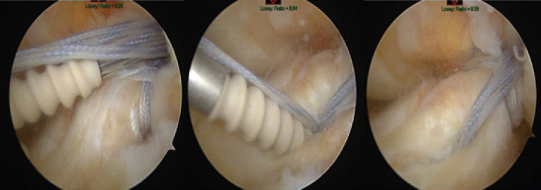
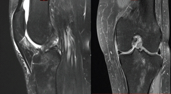
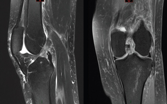
Case report: The authors present a case of a 60-year-old Caucasian female who developed a femoral-sided cyclops lesion after ACL repair. After initially achieving full range of motion (ROM) post-surgery, the patient later experienced a palpable clunk and extension loss 3-months postoperatively. While magnetic resonance imaging (MRI) was unremarkable, subsequent arthroscopy confirmed the diagnosis, leading to successful lesion excision and notchplasty.
Conclusion: A high index of suspicion for cyclops lesions is critical in patients presenting with a clunk with terminal extension after ACL repair, even in the absence of MRI evidence. Prompt recognition and intervention are crucial, as demonstrated by the removal of the lesion and notchplasty, which led to full recovery of symptoms and ROM of the knee. The authors aim to broaden the limited existing knowledge of inverted cyclops lesions by presenting a detailed case report of a patient after an ACL repair.

 求助内容:
求助内容: 应助结果提醒方式:


