{"title":"孤立的胸椎前硬膜外肠内生囊肿与胸前脑膜膨出相似:1例报告。","authors":"Surya Prakash Rao Voleti, Apurve Parameswaran, Ranjith Mahesh Nellore, Raja Shekar Kaitepalli, Vidya Kedarisetti","doi":"10.13107/jocr.2025.v15.i09.6014","DOIUrl":null,"url":null,"abstract":"<p><strong>Introduction: </strong>Enterogenous cysts (also known as neurenteric cysts) are rare congenital lesions presumed to arise from an abnormal persistent communication between embryonic ectodermal and endodermal tissues. They may be symptomatic or asymptomatic, and generally have an insidious and prolonged clinical course. They are typically intradural and extramedullary in location. Very few cases of extramedullary enterogenous cysts have been reported in the literature, most of which were cranial. We present a rare case of an anteriorly situated extradural enterogenous cyst at the level of the thoracic spine, mimicking an isolated anterior thoracic meningocele on radiologic evaluation.</p><p><strong>Case report: </strong>A 4-year-old boy was referred to our spine surgery unit for the evaluation and management of an incidentally detected T1 vertebral anomaly. Magnetic resonance imaging revealed the presence of a focal defect over the right half of the T1 vertebral body, through which herniation of a fluid-filled sac into the pre- and right paravertebral regions was noted. A diagnosis of anterior thoracic meningocele was made, and surgery was advised. Following surgical exposure of the entire extent of the sac, needle aspiration to decompress the lesion was performed, which yielded a milky-colored viscous fluid, unlike cerebrospinal fluid. The likelihood of an enterogenous cyst was suspected. The lesion was excised at the level of the base of its peduncle, and the vertebral defect was closed using a small contoured plate. Histopathologic evaluation confirmed the presence of an enterogenous cyst.</p><p><strong>Conclusion: </strong>Anterior thoracic meningoceles and enterogenous cysts of the spine may be indistinguishable on radiologic evaluation, and a definitive diagnosis may be reached only through histopathologic evaluation. The possibility of an enterogenous cyst must be considered in the differential diagnosis of meningoceles, and vice versa.</p>","PeriodicalId":16647,"journal":{"name":"Journal of Orthopaedic Case Reports","volume":"15 9","pages":"56-61"},"PeriodicalIF":0.0000,"publicationDate":"2025-09-01","publicationTypes":"Journal Article","fieldsOfStudy":null,"isOpenAccess":false,"openAccessPdf":"https://www.ncbi.nlm.nih.gov/pmc/articles/PMC12422650/pdf/","citationCount":"0","resultStr":"{\"title\":\"Isolated Anterior Extradural Enterogenous Cyst of the Thoracic Spine Mimicking an Anterior Thoracic Meningocele: A Case Report.\",\"authors\":\"Surya Prakash Rao Voleti, Apurve Parameswaran, Ranjith Mahesh Nellore, Raja Shekar Kaitepalli, Vidya Kedarisetti\",\"doi\":\"10.13107/jocr.2025.v15.i09.6014\",\"DOIUrl\":null,\"url\":null,\"abstract\":\"<p><strong>Introduction: </strong>Enterogenous cysts (also known as neurenteric cysts) are rare congenital lesions presumed to arise from an abnormal persistent communication between embryonic ectodermal and endodermal tissues. They may be symptomatic or asymptomatic, and generally have an insidious and prolonged clinical course. They are typically intradural and extramedullary in location. Very few cases of extramedullary enterogenous cysts have been reported in the literature, most of which were cranial. We present a rare case of an anteriorly situated extradural enterogenous cyst at the level of the thoracic spine, mimicking an isolated anterior thoracic meningocele on radiologic evaluation.</p><p><strong>Case report: </strong>A 4-year-old boy was referred to our spine surgery unit for the evaluation and management of an incidentally detected T1 vertebral anomaly. Magnetic resonance imaging revealed the presence of a focal defect over the right half of the T1 vertebral body, through which herniation of a fluid-filled sac into the pre- and right paravertebral regions was noted. A diagnosis of anterior thoracic meningocele was made, and surgery was advised. Following surgical exposure of the entire extent of the sac, needle aspiration to decompress the lesion was performed, which yielded a milky-colored viscous fluid, unlike cerebrospinal fluid. The likelihood of an enterogenous cyst was suspected. The lesion was excised at the level of the base of its peduncle, and the vertebral defect was closed using a small contoured plate. Histopathologic evaluation confirmed the presence of an enterogenous cyst.</p><p><strong>Conclusion: </strong>Anterior thoracic meningoceles and enterogenous cysts of the spine may be indistinguishable on radiologic evaluation, and a definitive diagnosis may be reached only through histopathologic evaluation. The possibility of an enterogenous cyst must be considered in the differential diagnosis of meningoceles, and vice versa.</p>\",\"PeriodicalId\":16647,\"journal\":{\"name\":\"Journal of Orthopaedic Case Reports\",\"volume\":\"15 9\",\"pages\":\"56-61\"},\"PeriodicalIF\":0.0000,\"publicationDate\":\"2025-09-01\",\"publicationTypes\":\"Journal Article\",\"fieldsOfStudy\":null,\"isOpenAccess\":false,\"openAccessPdf\":\"https://www.ncbi.nlm.nih.gov/pmc/articles/PMC12422650/pdf/\",\"citationCount\":\"0\",\"resultStr\":null,\"platform\":\"Semanticscholar\",\"paperid\":null,\"PeriodicalName\":\"Journal of Orthopaedic Case Reports\",\"FirstCategoryId\":\"1085\",\"ListUrlMain\":\"https://doi.org/10.13107/jocr.2025.v15.i09.6014\",\"RegionNum\":0,\"RegionCategory\":null,\"ArticlePicture\":[],\"TitleCN\":null,\"AbstractTextCN\":null,\"PMCID\":null,\"EPubDate\":\"\",\"PubModel\":\"\",\"JCR\":\"\",\"JCRName\":\"\",\"Score\":null,\"Total\":0}","platform":"Semanticscholar","paperid":null,"PeriodicalName":"Journal of Orthopaedic Case Reports","FirstCategoryId":"1085","ListUrlMain":"https://doi.org/10.13107/jocr.2025.v15.i09.6014","RegionNum":0,"RegionCategory":null,"ArticlePicture":[],"TitleCN":null,"AbstractTextCN":null,"PMCID":null,"EPubDate":"","PubModel":"","JCR":"","JCRName":"","Score":null,"Total":0}
Isolated Anterior Extradural Enterogenous Cyst of the Thoracic Spine Mimicking an Anterior Thoracic Meningocele: A Case Report.
Introduction: Enterogenous cysts (also known as neurenteric cysts) are rare congenital lesions presumed to arise from an abnormal persistent communication between embryonic ectodermal and endodermal tissues. They may be symptomatic or asymptomatic, and generally have an insidious and prolonged clinical course. They are typically intradural and extramedullary in location. Very few cases of extramedullary enterogenous cysts have been reported in the literature, most of which were cranial. We present a rare case of an anteriorly situated extradural enterogenous cyst at the level of the thoracic spine, mimicking an isolated anterior thoracic meningocele on radiologic evaluation.
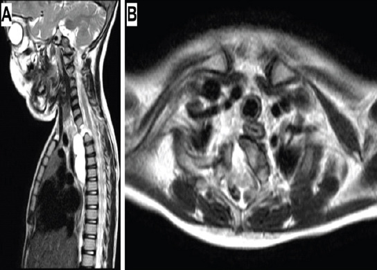
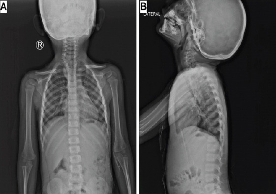
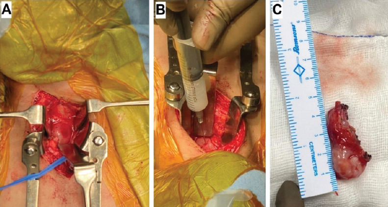
Case report: A 4-year-old boy was referred to our spine surgery unit for the evaluation and management of an incidentally detected T1 vertebral anomaly. Magnetic resonance imaging revealed the presence of a focal defect over the right half of the T1 vertebral body, through which herniation of a fluid-filled sac into the pre- and right paravertebral regions was noted. A diagnosis of anterior thoracic meningocele was made, and surgery was advised. Following surgical exposure of the entire extent of the sac, needle aspiration to decompress the lesion was performed, which yielded a milky-colored viscous fluid, unlike cerebrospinal fluid. The likelihood of an enterogenous cyst was suspected. The lesion was excised at the level of the base of its peduncle, and the vertebral defect was closed using a small contoured plate. Histopathologic evaluation confirmed the presence of an enterogenous cyst.
Conclusion: Anterior thoracic meningoceles and enterogenous cysts of the spine may be indistinguishable on radiologic evaluation, and a definitive diagnosis may be reached only through histopathologic evaluation. The possibility of an enterogenous cyst must be considered in the differential diagnosis of meningoceles, and vice versa.

 求助内容:
求助内容: 应助结果提醒方式:


