M S Karthik, Abrar Mohammed, Avinash Parthasarathy
{"title":"全膝关节置换术后异位骨化冲击股四头肌机制的罕见病例:一例报告。","authors":"M S Karthik, Abrar Mohammed, Avinash Parthasarathy","doi":"10.13107/jocr.2025.v15.i09.6024","DOIUrl":null,"url":null,"abstract":"<p><strong>Introduction: </strong>Heterotopic ossification (HO) is a rare complication following total knee replacement (TKR), with an incidence ranging between 1% and 3%. This condition can lead to significant functional limitations, including immobility and pain, particularly when ossification impinges on adjacent structures.</p><p><strong>Case report: </strong>We report the case of a 67-year-old female with grade 4 osteoarthritis of the left knee who underwent TKR following the failure of conservative management. Postoperatively, the patient developed stiffness and a reduced range of motion. Radiographic imaging at 5 weeks showed haziness in the anterior aspect of the femur at the upper border of the femoral implant, with gradual progression to distinct ossification by 3 months.</p><p><strong>Results: </strong>The heterotopic bone formation was found to impinge on the quadriceps mechanism, limiting knee flexion but not preventing full extension. Conservative management involving physiotherapy and analgesics was pursued, and surgical excision was avoided initially. Over 1 year, the patient showed improvement in extension, although no significant improvement in flexion was observed. Surgical excision of the heterotopic bone was performed at the end of 1 year, followed by post-operative radiotherapy to prevent recurrence.</p><p><strong>Conclusion: </strong>This case underscores the rarity of HO following TKR, particularly when it involves the quadriceps mechanism. It highlights the importance of early detection, close monitoring, and a stepwise treatment approach - starting with conservative therapy and escalating to surgical intervention when necessary. Post-operative radiotherapy can be considered in select cases to minimize recurrence.</p>","PeriodicalId":16647,"journal":{"name":"Journal of Orthopaedic Case Reports","volume":"15 9","pages":"83-87"},"PeriodicalIF":0.0000,"publicationDate":"2025-09-01","publicationTypes":"Journal Article","fieldsOfStudy":null,"isOpenAccess":false,"openAccessPdf":"https://www.ncbi.nlm.nih.gov/pmc/articles/PMC12422689/pdf/","citationCount":"0","resultStr":"{\"title\":\"Rare Case of Heterotopic Ossification Impinging on the Quadriceps Mechanism Following Total Knee Replacement: A Case Report.\",\"authors\":\"M S Karthik, Abrar Mohammed, Avinash Parthasarathy\",\"doi\":\"10.13107/jocr.2025.v15.i09.6024\",\"DOIUrl\":null,\"url\":null,\"abstract\":\"<p><strong>Introduction: </strong>Heterotopic ossification (HO) is a rare complication following total knee replacement (TKR), with an incidence ranging between 1% and 3%. This condition can lead to significant functional limitations, including immobility and pain, particularly when ossification impinges on adjacent structures.</p><p><strong>Case report: </strong>We report the case of a 67-year-old female with grade 4 osteoarthritis of the left knee who underwent TKR following the failure of conservative management. Postoperatively, the patient developed stiffness and a reduced range of motion. Radiographic imaging at 5 weeks showed haziness in the anterior aspect of the femur at the upper border of the femoral implant, with gradual progression to distinct ossification by 3 months.</p><p><strong>Results: </strong>The heterotopic bone formation was found to impinge on the quadriceps mechanism, limiting knee flexion but not preventing full extension. Conservative management involving physiotherapy and analgesics was pursued, and surgical excision was avoided initially. Over 1 year, the patient showed improvement in extension, although no significant improvement in flexion was observed. Surgical excision of the heterotopic bone was performed at the end of 1 year, followed by post-operative radiotherapy to prevent recurrence.</p><p><strong>Conclusion: </strong>This case underscores the rarity of HO following TKR, particularly when it involves the quadriceps mechanism. It highlights the importance of early detection, close monitoring, and a stepwise treatment approach - starting with conservative therapy and escalating to surgical intervention when necessary. Post-operative radiotherapy can be considered in select cases to minimize recurrence.</p>\",\"PeriodicalId\":16647,\"journal\":{\"name\":\"Journal of Orthopaedic Case Reports\",\"volume\":\"15 9\",\"pages\":\"83-87\"},\"PeriodicalIF\":0.0000,\"publicationDate\":\"2025-09-01\",\"publicationTypes\":\"Journal Article\",\"fieldsOfStudy\":null,\"isOpenAccess\":false,\"openAccessPdf\":\"https://www.ncbi.nlm.nih.gov/pmc/articles/PMC12422689/pdf/\",\"citationCount\":\"0\",\"resultStr\":null,\"platform\":\"Semanticscholar\",\"paperid\":null,\"PeriodicalName\":\"Journal of Orthopaedic Case Reports\",\"FirstCategoryId\":\"1085\",\"ListUrlMain\":\"https://doi.org/10.13107/jocr.2025.v15.i09.6024\",\"RegionNum\":0,\"RegionCategory\":null,\"ArticlePicture\":[],\"TitleCN\":null,\"AbstractTextCN\":null,\"PMCID\":null,\"EPubDate\":\"\",\"PubModel\":\"\",\"JCR\":\"\",\"JCRName\":\"\",\"Score\":null,\"Total\":0}","platform":"Semanticscholar","paperid":null,"PeriodicalName":"Journal of Orthopaedic Case Reports","FirstCategoryId":"1085","ListUrlMain":"https://doi.org/10.13107/jocr.2025.v15.i09.6024","RegionNum":0,"RegionCategory":null,"ArticlePicture":[],"TitleCN":null,"AbstractTextCN":null,"PMCID":null,"EPubDate":"","PubModel":"","JCR":"","JCRName":"","Score":null,"Total":0}
Rare Case of Heterotopic Ossification Impinging on the Quadriceps Mechanism Following Total Knee Replacement: A Case Report.
Introduction: Heterotopic ossification (HO) is a rare complication following total knee replacement (TKR), with an incidence ranging between 1% and 3%. This condition can lead to significant functional limitations, including immobility and pain, particularly when ossification impinges on adjacent structures.
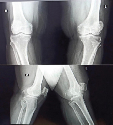
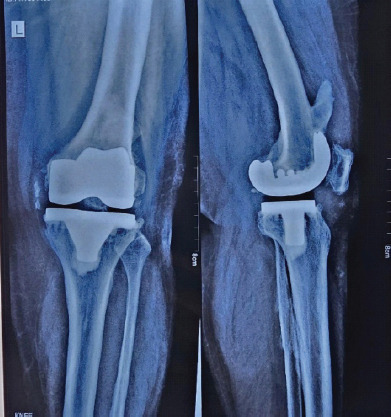
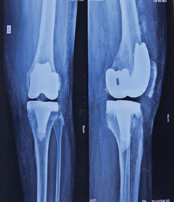
Case report: We report the case of a 67-year-old female with grade 4 osteoarthritis of the left knee who underwent TKR following the failure of conservative management. Postoperatively, the patient developed stiffness and a reduced range of motion. Radiographic imaging at 5 weeks showed haziness in the anterior aspect of the femur at the upper border of the femoral implant, with gradual progression to distinct ossification by 3 months.
Results: The heterotopic bone formation was found to impinge on the quadriceps mechanism, limiting knee flexion but not preventing full extension. Conservative management involving physiotherapy and analgesics was pursued, and surgical excision was avoided initially. Over 1 year, the patient showed improvement in extension, although no significant improvement in flexion was observed. Surgical excision of the heterotopic bone was performed at the end of 1 year, followed by post-operative radiotherapy to prevent recurrence.
Conclusion: This case underscores the rarity of HO following TKR, particularly when it involves the quadriceps mechanism. It highlights the importance of early detection, close monitoring, and a stepwise treatment approach - starting with conservative therapy and escalating to surgical intervention when necessary. Post-operative radiotherapy can be considered in select cases to minimize recurrence.

 求助内容:
求助内容: 应助结果提醒方式:


