{"title":"牛津单室膝关节置换术后罕见的假体周围骨折:股骨髁上和胫骨近端骨折病例系列。","authors":"Amyn M Rajani, Vishal Kulkarni, Clevio Desouza","doi":"10.13107/jocr.2025.v15.i09.6054","DOIUrl":null,"url":null,"abstract":"<p><strong>Introduction: </strong>Periprosthetic fractures (PPFs) are well-documented complications after total knee arthroplasty but are exceedingly rare following unicompartmental knee arthroplasty (UKA). Given UKA's design preserves native bone stock and maintains near-normal joint biomechanics, PPFs around well-fixed UKA components are infrequently encountered and poorly described in the literature. To the best of our knowledge, there are very few detailed reports of supracondylar femoral or proximal tibial PPFs following mobile-bearing UKA. This case series is important because it highlights not only the rarity of these injuries but also their successful management without compromising the integrity of the prosthetic components.</p><p><strong>Case report: </strong>We present two unique cases of PPFs following cemented mobile-bearing Oxford UKA in elderly South Asian women. The first case involves a 63-year-old woman who sustained a high-energy supracondylar femoral PPF (Unified Classification System Type C) 9 months post-UKA. Despite the severity of the injury, radiographs confirmed the femoral component remained well-fixed with no evidence of polyethylene insert dislocation. She was treated successfully with retrograde intramedullary nailing, achieving full fracture union and 130° of knee flexion by 6 months postoperatively. The second case involves a 60-year-old woman who sustained a proximal third tibial PPF (Type C) two and a half months after UKA. Again, both components remained secure without signs of loosening. She was treated with locking plate fixation, resulting in complete union and full independent ambulation by 6 months. Both patients remained clinically well at 2.5 years of follow-up, with intact UKA components and no functional limitations.</p><p><strong>Conclusion: </strong>These cases underscore that even severe, high-energy PPFs around UKA can be effectively managed with standard anatomical fixation techniques without necessitating implant revision. The report advances clinical knowledge by demonstrating the structural resilience of well-fixed mobile-bearing UKA components in the setting of traumatic fracture. It is of particular interest to orthopaedic surgeons specializing in arthroplasty and trauma, but also has broader implications for surgical planning, patient counseling, and post-operative expectations.</p>","PeriodicalId":16647,"journal":{"name":"Journal of Orthopaedic Case Reports","volume":"15 9","pages":"164-169"},"PeriodicalIF":0.0000,"publicationDate":"2025-09-01","publicationTypes":"Journal Article","fieldsOfStudy":null,"isOpenAccess":false,"openAccessPdf":"https://www.ncbi.nlm.nih.gov/pmc/articles/PMC12422644/pdf/","citationCount":"0","resultStr":"{\"title\":\"Rare Periprosthetic Fractures Following Oxford Unicompartmental Knee Arthroplasty: A Case Series of Supracondylar Femoral and Proximal Tibial Fractures.\",\"authors\":\"Amyn M Rajani, Vishal Kulkarni, Clevio Desouza\",\"doi\":\"10.13107/jocr.2025.v15.i09.6054\",\"DOIUrl\":null,\"url\":null,\"abstract\":\"<p><strong>Introduction: </strong>Periprosthetic fractures (PPFs) are well-documented complications after total knee arthroplasty but are exceedingly rare following unicompartmental knee arthroplasty (UKA). Given UKA's design preserves native bone stock and maintains near-normal joint biomechanics, PPFs around well-fixed UKA components are infrequently encountered and poorly described in the literature. To the best of our knowledge, there are very few detailed reports of supracondylar femoral or proximal tibial PPFs following mobile-bearing UKA. This case series is important because it highlights not only the rarity of these injuries but also their successful management without compromising the integrity of the prosthetic components.</p><p><strong>Case report: </strong>We present two unique cases of PPFs following cemented mobile-bearing Oxford UKA in elderly South Asian women. The first case involves a 63-year-old woman who sustained a high-energy supracondylar femoral PPF (Unified Classification System Type C) 9 months post-UKA. Despite the severity of the injury, radiographs confirmed the femoral component remained well-fixed with no evidence of polyethylene insert dislocation. She was treated successfully with retrograde intramedullary nailing, achieving full fracture union and 130° of knee flexion by 6 months postoperatively. The second case involves a 60-year-old woman who sustained a proximal third tibial PPF (Type C) two and a half months after UKA. Again, both components remained secure without signs of loosening. She was treated with locking plate fixation, resulting in complete union and full independent ambulation by 6 months. Both patients remained clinically well at 2.5 years of follow-up, with intact UKA components and no functional limitations.</p><p><strong>Conclusion: </strong>These cases underscore that even severe, high-energy PPFs around UKA can be effectively managed with standard anatomical fixation techniques without necessitating implant revision. The report advances clinical knowledge by demonstrating the structural resilience of well-fixed mobile-bearing UKA components in the setting of traumatic fracture. It is of particular interest to orthopaedic surgeons specializing in arthroplasty and trauma, but also has broader implications for surgical planning, patient counseling, and post-operative expectations.</p>\",\"PeriodicalId\":16647,\"journal\":{\"name\":\"Journal of Orthopaedic Case Reports\",\"volume\":\"15 9\",\"pages\":\"164-169\"},\"PeriodicalIF\":0.0000,\"publicationDate\":\"2025-09-01\",\"publicationTypes\":\"Journal Article\",\"fieldsOfStudy\":null,\"isOpenAccess\":false,\"openAccessPdf\":\"https://www.ncbi.nlm.nih.gov/pmc/articles/PMC12422644/pdf/\",\"citationCount\":\"0\",\"resultStr\":null,\"platform\":\"Semanticscholar\",\"paperid\":null,\"PeriodicalName\":\"Journal of Orthopaedic Case Reports\",\"FirstCategoryId\":\"1085\",\"ListUrlMain\":\"https://doi.org/10.13107/jocr.2025.v15.i09.6054\",\"RegionNum\":0,\"RegionCategory\":null,\"ArticlePicture\":[],\"TitleCN\":null,\"AbstractTextCN\":null,\"PMCID\":null,\"EPubDate\":\"\",\"PubModel\":\"\",\"JCR\":\"\",\"JCRName\":\"\",\"Score\":null,\"Total\":0}","platform":"Semanticscholar","paperid":null,"PeriodicalName":"Journal of Orthopaedic Case Reports","FirstCategoryId":"1085","ListUrlMain":"https://doi.org/10.13107/jocr.2025.v15.i09.6054","RegionNum":0,"RegionCategory":null,"ArticlePicture":[],"TitleCN":null,"AbstractTextCN":null,"PMCID":null,"EPubDate":"","PubModel":"","JCR":"","JCRName":"","Score":null,"Total":0}
Rare Periprosthetic Fractures Following Oxford Unicompartmental Knee Arthroplasty: A Case Series of Supracondylar Femoral and Proximal Tibial Fractures.
Introduction: Periprosthetic fractures (PPFs) are well-documented complications after total knee arthroplasty but are exceedingly rare following unicompartmental knee arthroplasty (UKA). Given UKA's design preserves native bone stock and maintains near-normal joint biomechanics, PPFs around well-fixed UKA components are infrequently encountered and poorly described in the literature. To the best of our knowledge, there are very few detailed reports of supracondylar femoral or proximal tibial PPFs following mobile-bearing UKA. This case series is important because it highlights not only the rarity of these injuries but also their successful management without compromising the integrity of the prosthetic components.
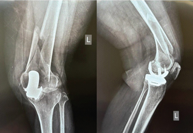
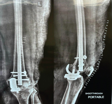
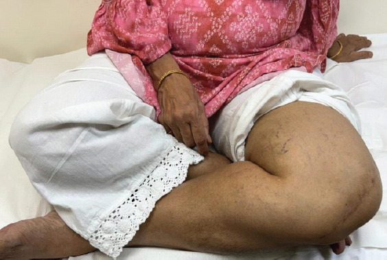
Case report: We present two unique cases of PPFs following cemented mobile-bearing Oxford UKA in elderly South Asian women. The first case involves a 63-year-old woman who sustained a high-energy supracondylar femoral PPF (Unified Classification System Type C) 9 months post-UKA. Despite the severity of the injury, radiographs confirmed the femoral component remained well-fixed with no evidence of polyethylene insert dislocation. She was treated successfully with retrograde intramedullary nailing, achieving full fracture union and 130° of knee flexion by 6 months postoperatively. The second case involves a 60-year-old woman who sustained a proximal third tibial PPF (Type C) two and a half months after UKA. Again, both components remained secure without signs of loosening. She was treated with locking plate fixation, resulting in complete union and full independent ambulation by 6 months. Both patients remained clinically well at 2.5 years of follow-up, with intact UKA components and no functional limitations.
Conclusion: These cases underscore that even severe, high-energy PPFs around UKA can be effectively managed with standard anatomical fixation techniques without necessitating implant revision. The report advances clinical knowledge by demonstrating the structural resilience of well-fixed mobile-bearing UKA components in the setting of traumatic fracture. It is of particular interest to orthopaedic surgeons specializing in arthroplasty and trauma, but also has broader implications for surgical planning, patient counseling, and post-operative expectations.

 求助内容:
求助内容: 应助结果提醒方式:


