Elizabeth Jelpke, Saphalya Pattnaik, Gur Aziz Sidhu
{"title":"跟骨与创伤性应力性骨折。","authors":"Elizabeth Jelpke, Saphalya Pattnaik, Gur Aziz Sidhu","doi":"10.13107/jocr.2025.v15.i09.6062","DOIUrl":null,"url":null,"abstract":"<p><strong>Introduction: </strong>Although uncommon, the calcaneus stress fracture is an important differential diagnosis of both traumatic and non-traumatic foot pain. The calcaneus is one of the tarsal bones that are prone to stress fractures, which usually occur as a result of overuse. The diagnosis of stress fractures is aided by plain radiographs, with the mainstay of management usually conservative.</p><p><strong>Case report: </strong>This case report is of a 57-year-old female who presented with instant left-sided heel pain after stepping off a step at home. Investigations included plain radiographs of the left foot and ankle, with no obvious fractures visible. As a result, a magnetic resonance imaging was obtained, which confirmed a stress fracture of the left os calcis. Management remained conservative, with the patient placed in an ankle boot for 4-6 weeks with non-weight bearing instructions provided. Heel pain can be caused by a stress fracture of the calcaneus, and although these injuries are usually caused by repetitive forces, this case study provides a reminder that they can also be caused by acute trauma.</p><p><strong>Conclusion: </strong>Calcaneal stress fractures, typically due to overuse, can also result from acute trauma and may require MRI for diagnosis when plain radiographs are inconclusive. The mainstay of treatment is conservative management.</p>","PeriodicalId":16647,"journal":{"name":"Journal of Orthopaedic Case Reports","volume":"15 9","pages":"185-187"},"PeriodicalIF":0.0000,"publicationDate":"2025-09-01","publicationTypes":"Journal Article","fieldsOfStudy":null,"isOpenAccess":false,"openAccessPdf":"https://www.ncbi.nlm.nih.gov/pmc/articles/PMC12422668/pdf/","citationCount":"0","resultStr":"{\"title\":\"Calcaneus and Traumatic Stress Fracture.\",\"authors\":\"Elizabeth Jelpke, Saphalya Pattnaik, Gur Aziz Sidhu\",\"doi\":\"10.13107/jocr.2025.v15.i09.6062\",\"DOIUrl\":null,\"url\":null,\"abstract\":\"<p><strong>Introduction: </strong>Although uncommon, the calcaneus stress fracture is an important differential diagnosis of both traumatic and non-traumatic foot pain. The calcaneus is one of the tarsal bones that are prone to stress fractures, which usually occur as a result of overuse. The diagnosis of stress fractures is aided by plain radiographs, with the mainstay of management usually conservative.</p><p><strong>Case report: </strong>This case report is of a 57-year-old female who presented with instant left-sided heel pain after stepping off a step at home. Investigations included plain radiographs of the left foot and ankle, with no obvious fractures visible. As a result, a magnetic resonance imaging was obtained, which confirmed a stress fracture of the left os calcis. Management remained conservative, with the patient placed in an ankle boot for 4-6 weeks with non-weight bearing instructions provided. Heel pain can be caused by a stress fracture of the calcaneus, and although these injuries are usually caused by repetitive forces, this case study provides a reminder that they can also be caused by acute trauma.</p><p><strong>Conclusion: </strong>Calcaneal stress fractures, typically due to overuse, can also result from acute trauma and may require MRI for diagnosis when plain radiographs are inconclusive. The mainstay of treatment is conservative management.</p>\",\"PeriodicalId\":16647,\"journal\":{\"name\":\"Journal of Orthopaedic Case Reports\",\"volume\":\"15 9\",\"pages\":\"185-187\"},\"PeriodicalIF\":0.0000,\"publicationDate\":\"2025-09-01\",\"publicationTypes\":\"Journal Article\",\"fieldsOfStudy\":null,\"isOpenAccess\":false,\"openAccessPdf\":\"https://www.ncbi.nlm.nih.gov/pmc/articles/PMC12422668/pdf/\",\"citationCount\":\"0\",\"resultStr\":null,\"platform\":\"Semanticscholar\",\"paperid\":null,\"PeriodicalName\":\"Journal of Orthopaedic Case Reports\",\"FirstCategoryId\":\"1085\",\"ListUrlMain\":\"https://doi.org/10.13107/jocr.2025.v15.i09.6062\",\"RegionNum\":0,\"RegionCategory\":null,\"ArticlePicture\":[],\"TitleCN\":null,\"AbstractTextCN\":null,\"PMCID\":null,\"EPubDate\":\"\",\"PubModel\":\"\",\"JCR\":\"\",\"JCRName\":\"\",\"Score\":null,\"Total\":0}","platform":"Semanticscholar","paperid":null,"PeriodicalName":"Journal of Orthopaedic Case Reports","FirstCategoryId":"1085","ListUrlMain":"https://doi.org/10.13107/jocr.2025.v15.i09.6062","RegionNum":0,"RegionCategory":null,"ArticlePicture":[],"TitleCN":null,"AbstractTextCN":null,"PMCID":null,"EPubDate":"","PubModel":"","JCR":"","JCRName":"","Score":null,"Total":0}
Introduction: Although uncommon, the calcaneus stress fracture is an important differential diagnosis of both traumatic and non-traumatic foot pain. The calcaneus is one of the tarsal bones that are prone to stress fractures, which usually occur as a result of overuse. The diagnosis of stress fractures is aided by plain radiographs, with the mainstay of management usually conservative.
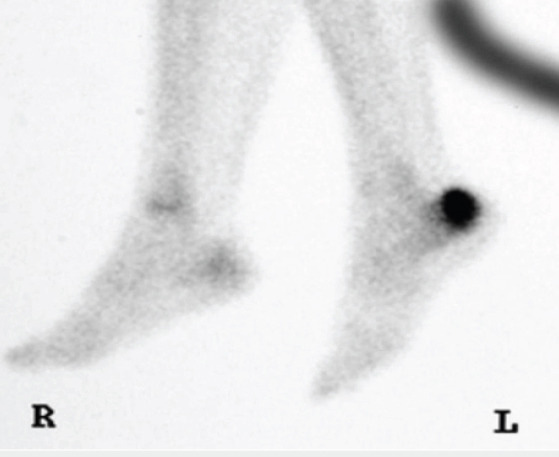
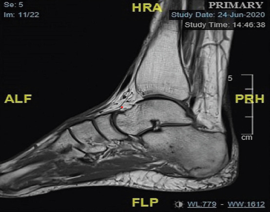
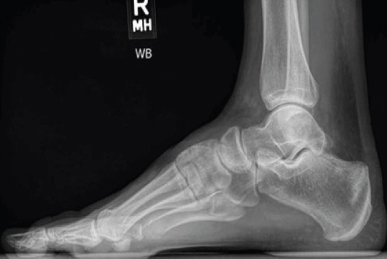
Case report: This case report is of a 57-year-old female who presented with instant left-sided heel pain after stepping off a step at home. Investigations included plain radiographs of the left foot and ankle, with no obvious fractures visible. As a result, a magnetic resonance imaging was obtained, which confirmed a stress fracture of the left os calcis. Management remained conservative, with the patient placed in an ankle boot for 4-6 weeks with non-weight bearing instructions provided. Heel pain can be caused by a stress fracture of the calcaneus, and although these injuries are usually caused by repetitive forces, this case study provides a reminder that they can also be caused by acute trauma.
Conclusion: Calcaneal stress fractures, typically due to overuse, can also result from acute trauma and may require MRI for diagnosis when plain radiographs are inconclusive. The mainstay of treatment is conservative management.

 求助内容:
求助内容: 应助结果提醒方式:


