Anna Sosa, Kimberly A O'Neill, Ruben Jauregui, Ugo Nwigwe, Thibo Billiet, Rachel Kenney, Lauren B Krupp, Steven L Galetta, Laura J Balcer, Scott N Grossman
{"title":"小儿多发性硬化症视功能、视网膜厚度与MRI脑容量的关系。","authors":"Anna Sosa, Kimberly A O'Neill, Ruben Jauregui, Ugo Nwigwe, Thibo Billiet, Rachel Kenney, Lauren B Krupp, Steven L Galetta, Laura J Balcer, Scott N Grossman","doi":"10.1212/NXI.0000000000200480","DOIUrl":null,"url":null,"abstract":"<p><strong>Background and objectives: </strong>While reductions in optical coherence tomography (OCT) pRNFL and ganglion cell-inner plexiform layer thicknesses have been shown to be associated with brain atrophy in adult-onset MS (AOMS) cohorts, the relationship between OCT and brain MRI measures is less established in pediatric-onset MS (POMS). Our aim was to examine the associations of OCT measures with volumetric MRI in a cohort of patients with POMS to determine whether OCT measures reflect CNS neurodegeneration in this patient population, as is seen in AOMS cohorts.</p><p><strong>Methods: </strong>This was a cross-sectional study with retrospective ascertainment of patients with POMS evaluated at a single center with expertise in POMS and neuro-ophthalmology. As part of routine clinical care, patients with POMS are evaluated by a POMS expert and undergo volumetric brain MRI, including whole-brain (WB), subregional, and gray matter (GM) volume analyses. Patients with POMS are routinely referred to neuro-ophthalmology for evaluation that includes high-contrast visual acuity, color vision testing, and OCT. Generalized estimating equation (GEE) models, accounting for within-patient, intereye correlations (both eyes of each patient were included), MS disease duration, and disease-modifying therapy efficacy, were used to determine the relationship between visual pathway structure and function and volumetric MRI measures.</p><p><strong>Results: </strong>Among 61 patients (122 eyes) with POMS, the mean age at the time of the volumetric MRI scan was 19.2 (SD = 3.7, range 10-27) years, with a median disease duration of 2.8 (range 0-14) years. Lower (worse) pRNFL thicknesses (mean 87.4 [17.2] µm) were associated with reduced volume percentiles of WB (<i>p</i> < 0.001, GEE models), total GM (<i>p</i> = 0.025), and thalamus (<i>p</i> = 0.038). pRNFL thinning was also associated with greater lesion (<i>p</i> = 0.006) and black hole (<i>p</i> = 0.028) volumes. Reduced color vision and decreased high-contrast visual acuity were associated with lower hippocampal volumes (<i>p</i> = 0.012 and <i>p</i> = 0.015, respectively).</p><p><strong>Discussion: </strong>Our results demonstrate that changes in visual pathway structures are associated with reductions in overall brain volume and GM volumes, as well as greater lesion and black hole burden. Collectively, our results emphasize the importance of visual assessment in POMS and suggest that OCT reflects overall CNS neurodegeneration in this cohort.</p>","PeriodicalId":19472,"journal":{"name":"Neurology® Neuroimmunology & Neuroinflammation","volume":"12 6","pages":"e200480"},"PeriodicalIF":7.5000,"publicationDate":"2025-11-01","publicationTypes":"Journal Article","fieldsOfStudy":null,"isOpenAccess":false,"openAccessPdf":"https://www.ncbi.nlm.nih.gov/pmc/articles/PMC12424074/pdf/","citationCount":"0","resultStr":"{\"title\":\"Relation of Visual Function, Retinal Thickness by Optical Coherence Tomography, and MRI Brain Volume in Pediatric-Onset Multiple Sclerosis.\",\"authors\":\"Anna Sosa, Kimberly A O'Neill, Ruben Jauregui, Ugo Nwigwe, Thibo Billiet, Rachel Kenney, Lauren B Krupp, Steven L Galetta, Laura J Balcer, Scott N Grossman\",\"doi\":\"10.1212/NXI.0000000000200480\",\"DOIUrl\":null,\"url\":null,\"abstract\":\"<p><strong>Background and objectives: </strong>While reductions in optical coherence tomography (OCT) pRNFL and ganglion cell-inner plexiform layer thicknesses have been shown to be associated with brain atrophy in adult-onset MS (AOMS) cohorts, the relationship between OCT and brain MRI measures is less established in pediatric-onset MS (POMS). Our aim was to examine the associations of OCT measures with volumetric MRI in a cohort of patients with POMS to determine whether OCT measures reflect CNS neurodegeneration in this patient population, as is seen in AOMS cohorts.</p><p><strong>Methods: </strong>This was a cross-sectional study with retrospective ascertainment of patients with POMS evaluated at a single center with expertise in POMS and neuro-ophthalmology. As part of routine clinical care, patients with POMS are evaluated by a POMS expert and undergo volumetric brain MRI, including whole-brain (WB), subregional, and gray matter (GM) volume analyses. Patients with POMS are routinely referred to neuro-ophthalmology for evaluation that includes high-contrast visual acuity, color vision testing, and OCT. Generalized estimating equation (GEE) models, accounting for within-patient, intereye correlations (both eyes of each patient were included), MS disease duration, and disease-modifying therapy efficacy, were used to determine the relationship between visual pathway structure and function and volumetric MRI measures.</p><p><strong>Results: </strong>Among 61 patients (122 eyes) with POMS, the mean age at the time of the volumetric MRI scan was 19.2 (SD = 3.7, range 10-27) years, with a median disease duration of 2.8 (range 0-14) years. Lower (worse) pRNFL thicknesses (mean 87.4 [17.2] µm) were associated with reduced volume percentiles of WB (<i>p</i> < 0.001, GEE models), total GM (<i>p</i> = 0.025), and thalamus (<i>p</i> = 0.038). pRNFL thinning was also associated with greater lesion (<i>p</i> = 0.006) and black hole (<i>p</i> = 0.028) volumes. Reduced color vision and decreased high-contrast visual acuity were associated with lower hippocampal volumes (<i>p</i> = 0.012 and <i>p</i> = 0.015, respectively).</p><p><strong>Discussion: </strong>Our results demonstrate that changes in visual pathway structures are associated with reductions in overall brain volume and GM volumes, as well as greater lesion and black hole burden. Collectively, our results emphasize the importance of visual assessment in POMS and suggest that OCT reflects overall CNS neurodegeneration in this cohort.</p>\",\"PeriodicalId\":19472,\"journal\":{\"name\":\"Neurology® Neuroimmunology & Neuroinflammation\",\"volume\":\"12 6\",\"pages\":\"e200480\"},\"PeriodicalIF\":7.5000,\"publicationDate\":\"2025-11-01\",\"publicationTypes\":\"Journal Article\",\"fieldsOfStudy\":null,\"isOpenAccess\":false,\"openAccessPdf\":\"https://www.ncbi.nlm.nih.gov/pmc/articles/PMC12424074/pdf/\",\"citationCount\":\"0\",\"resultStr\":null,\"platform\":\"Semanticscholar\",\"paperid\":null,\"PeriodicalName\":\"Neurology® Neuroimmunology & Neuroinflammation\",\"FirstCategoryId\":\"3\",\"ListUrlMain\":\"https://doi.org/10.1212/NXI.0000000000200480\",\"RegionNum\":1,\"RegionCategory\":\"医学\",\"ArticlePicture\":[],\"TitleCN\":null,\"AbstractTextCN\":null,\"PMCID\":null,\"EPubDate\":\"2025/9/9 0:00:00\",\"PubModel\":\"Epub\",\"JCR\":\"Q1\",\"JCRName\":\"CLINICAL NEUROLOGY\",\"Score\":null,\"Total\":0}","platform":"Semanticscholar","paperid":null,"PeriodicalName":"Neurology® Neuroimmunology & Neuroinflammation","FirstCategoryId":"3","ListUrlMain":"https://doi.org/10.1212/NXI.0000000000200480","RegionNum":1,"RegionCategory":"医学","ArticlePicture":[],"TitleCN":null,"AbstractTextCN":null,"PMCID":null,"EPubDate":"2025/9/9 0:00:00","PubModel":"Epub","JCR":"Q1","JCRName":"CLINICAL NEUROLOGY","Score":null,"Total":0}
Relation of Visual Function, Retinal Thickness by Optical Coherence Tomography, and MRI Brain Volume in Pediatric-Onset Multiple Sclerosis.
Background and objectives: While reductions in optical coherence tomography (OCT) pRNFL and ganglion cell-inner plexiform layer thicknesses have been shown to be associated with brain atrophy in adult-onset MS (AOMS) cohorts, the relationship between OCT and brain MRI measures is less established in pediatric-onset MS (POMS). Our aim was to examine the associations of OCT measures with volumetric MRI in a cohort of patients with POMS to determine whether OCT measures reflect CNS neurodegeneration in this patient population, as is seen in AOMS cohorts.
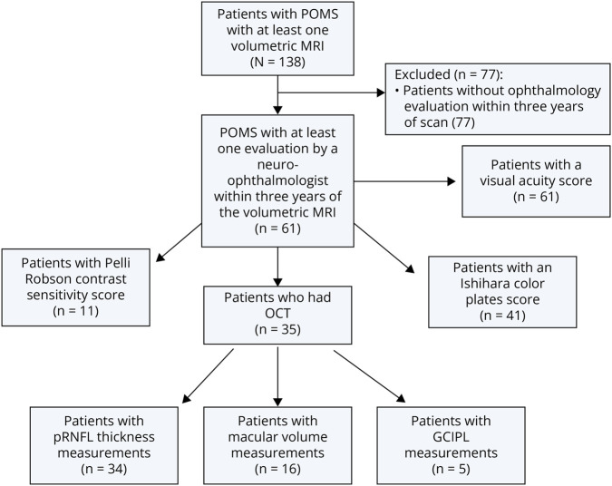
Methods: This was a cross-sectional study with retrospective ascertainment of patients with POMS evaluated at a single center with expertise in POMS and neuro-ophthalmology. As part of routine clinical care, patients with POMS are evaluated by a POMS expert and undergo volumetric brain MRI, including whole-brain (WB), subregional, and gray matter (GM) volume analyses. Patients with POMS are routinely referred to neuro-ophthalmology for evaluation that includes high-contrast visual acuity, color vision testing, and OCT. Generalized estimating equation (GEE) models, accounting for within-patient, intereye correlations (both eyes of each patient were included), MS disease duration, and disease-modifying therapy efficacy, were used to determine the relationship between visual pathway structure and function and volumetric MRI measures.
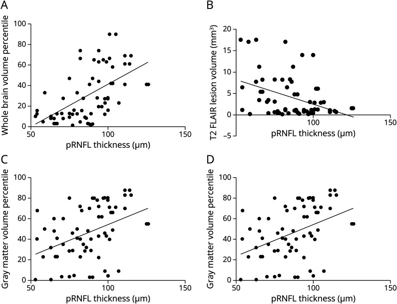
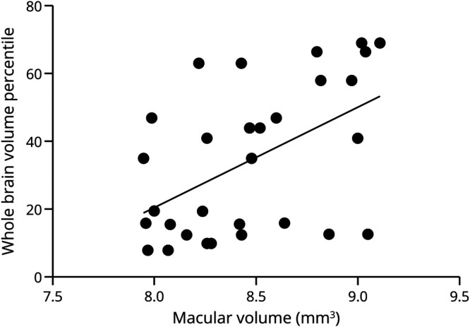
Results: Among 61 patients (122 eyes) with POMS, the mean age at the time of the volumetric MRI scan was 19.2 (SD = 3.7, range 10-27) years, with a median disease duration of 2.8 (range 0-14) years. Lower (worse) pRNFL thicknesses (mean 87.4 [17.2] µm) were associated with reduced volume percentiles of WB (p < 0.001, GEE models), total GM (p = 0.025), and thalamus (p = 0.038). pRNFL thinning was also associated with greater lesion (p = 0.006) and black hole (p = 0.028) volumes. Reduced color vision and decreased high-contrast visual acuity were associated with lower hippocampal volumes (p = 0.012 and p = 0.015, respectively).
Discussion: Our results demonstrate that changes in visual pathway structures are associated with reductions in overall brain volume and GM volumes, as well as greater lesion and black hole burden. Collectively, our results emphasize the importance of visual assessment in POMS and suggest that OCT reflects overall CNS neurodegeneration in this cohort.
期刊介绍:
Neurology Neuroimmunology & Neuroinflammation is an official journal of the American Academy of Neurology. Neurology: Neuroimmunology & Neuroinflammation will be the premier peer-reviewed journal in neuroimmunology and neuroinflammation. This journal publishes rigorously peer-reviewed open-access reports of original research and in-depth reviews of topics in neuroimmunology & neuroinflammation, affecting the full range of neurologic diseases including (but not limited to) Alzheimer's disease, Parkinson's disease, ALS, tauopathy, and stroke; multiple sclerosis and NMO; inflammatory peripheral nerve and muscle disease, Guillain-Barré and myasthenia gravis; nervous system infection; paraneoplastic syndromes, noninfectious encephalitides and other antibody-mediated disorders; and psychiatric and neurodevelopmental disorders. Clinical trials, instructive case reports, and small case series will also be featured.
 求助内容:
求助内容: 应助结果提醒方式:


