乳腺肿瘤的超声图像检测与解剖学的先验知识

IF 4.2 3区 计算机科学 Q2 COMPUTER SCIENCE, ARTIFICIAL INTELLIGENCE
Liangduan Wu , Yan Zhuang , Guoliang Liao , Lin Han , Zhan Hua , Rui Wang , Ke Chen , Jiangli Lin
{"title":"乳腺肿瘤的超声图像检测与解剖学的先验知识","authors":"Liangduan Wu ,&nbsp;Yan Zhuang ,&nbsp;Guoliang Liao ,&nbsp;Lin Han ,&nbsp;Zhan Hua ,&nbsp;Rui Wang ,&nbsp;Ke Chen ,&nbsp;Jiangli Lin","doi":"10.1016/j.imavis.2025.105724","DOIUrl":null,"url":null,"abstract":"<div><div>Breast tumor detection is an important step in the procedure of computer-aided diagnosis. In clinical practice, computer-aided diagnosis system not only processes lesion images but also processes normal images without lesions. However, normal images are often overlooked. In this study, we additionally collected numerous normal images to evaluate object detection algorithms. We found that similarities between tumors and hypoechoic regions have led to false positive lesions, and the frequency of false positive lesions in normal images is higher than it in lesion images. To address this issue, we incorporate anatomical prior knowledge of breast tumors to propose a novel breast tumor detection method. Our method consists of a preprocessing method and a novel breast tumor detection network. The preprocessing method automatically extracts breast regions as anatomical constraints and utilizes channel fusion to combine images of breast regions with original images. The proposed breast tumor detection network is based on programmable gradient information and large-kernel convolution. The programmable gradient information is applied by an auxiliary branch which provides more comprehensive gradient information for backpropagation, while large-kernel convolution expands the receptive field of neurons. As a result, our method achieves the best false positive lesion rate of 3.30% and gets a reduction by at least 5.67% over other compared algorithms in normal images, with the best precision of 90.91%, sensitivity of 88.57%, f1-score of 89.75%, and mean average precision of 93.07% in lesion images. Experimental results suggest promising application potential.</div></div>","PeriodicalId":50374,"journal":{"name":"Image and Vision Computing","volume":"162 ","pages":"Article 105724"},"PeriodicalIF":4.2000,"publicationDate":"2025-09-04","publicationTypes":"Journal Article","fieldsOfStudy":null,"isOpenAccess":false,"openAccessPdf":"","citationCount":"0","resultStr":"{\"title\":\"Breast tumor detection in ultrasound images with anatomical prior knowledge\",\"authors\":\"Liangduan Wu ,&nbsp;Yan Zhuang ,&nbsp;Guoliang Liao ,&nbsp;Lin Han ,&nbsp;Zhan Hua ,&nbsp;Rui Wang ,&nbsp;Ke Chen ,&nbsp;Jiangli Lin\",\"doi\":\"10.1016/j.imavis.2025.105724\",\"DOIUrl\":null,\"url\":null,\"abstract\":\"<div><div>Breast tumor detection is an important step in the procedure of computer-aided diagnosis. In clinical practice, computer-aided diagnosis system not only processes lesion images but also processes normal images without lesions. However, normal images are often overlooked. In this study, we additionally collected numerous normal images to evaluate object detection algorithms. We found that similarities between tumors and hypoechoic regions have led to false positive lesions, and the frequency of false positive lesions in normal images is higher than it in lesion images. To address this issue, we incorporate anatomical prior knowledge of breast tumors to propose a novel breast tumor detection method. Our method consists of a preprocessing method and a novel breast tumor detection network. The preprocessing method automatically extracts breast regions as anatomical constraints and utilizes channel fusion to combine images of breast regions with original images. The proposed breast tumor detection network is based on programmable gradient information and large-kernel convolution. The programmable gradient information is applied by an auxiliary branch which provides more comprehensive gradient information for backpropagation, while large-kernel convolution expands the receptive field of neurons. As a result, our method achieves the best false positive lesion rate of 3.30% and gets a reduction by at least 5.67% over other compared algorithms in normal images, with the best precision of 90.91%, sensitivity of 88.57%, f1-score of 89.75%, and mean average precision of 93.07% in lesion images. Experimental results suggest promising application potential.</div></div>\",\"PeriodicalId\":50374,\"journal\":{\"name\":\"Image and Vision Computing\",\"volume\":\"162 \",\"pages\":\"Article 105724\"},\"PeriodicalIF\":4.2000,\"publicationDate\":\"2025-09-04\",\"publicationTypes\":\"Journal Article\",\"fieldsOfStudy\":null,\"isOpenAccess\":false,\"openAccessPdf\":\"\",\"citationCount\":\"0\",\"resultStr\":null,\"platform\":\"Semanticscholar\",\"paperid\":null,\"PeriodicalName\":\"Image and Vision Computing\",\"FirstCategoryId\":\"94\",\"ListUrlMain\":\"https://www.sciencedirect.com/science/article/pii/S0262885625003129\",\"RegionNum\":3,\"RegionCategory\":\"计算机科学\",\"ArticlePicture\":[],\"TitleCN\":null,\"AbstractTextCN\":null,\"PMCID\":null,\"EPubDate\":\"\",\"PubModel\":\"\",\"JCR\":\"Q2\",\"JCRName\":\"COMPUTER SCIENCE, ARTIFICIAL INTELLIGENCE\",\"Score\":null,\"Total\":0}","platform":"Semanticscholar","paperid":null,"PeriodicalName":"Image and Vision Computing","FirstCategoryId":"94","ListUrlMain":"https://www.sciencedirect.com/science/article/pii/S0262885625003129","RegionNum":3,"RegionCategory":"计算机科学","ArticlePicture":[],"TitleCN":null,"AbstractTextCN":null,"PMCID":null,"EPubDate":"","PubModel":"","JCR":"Q2","JCRName":"COMPUTER SCIENCE, ARTIFICIAL INTELLIGENCE","Score":null,"Total":0}
引用次数: 0

摘要

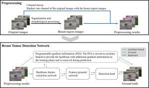
乳腺肿瘤的检测是计算机辅助诊断的重要步骤。在临床实践中,计算机辅助诊断系统不仅可以处理病变图像,还可以处理无病变的正常图像。然而,正常的图像往往被忽视。在这项研究中,我们还收集了大量的正常图像来评估目标检测算法。我们发现肿瘤与低回声区域之间的相似性导致了假阳性病变,并且正常图像中假阳性病变的频率高于病变图像。为了解决这一问题,我们结合乳腺肿瘤的解剖学先验知识,提出了一种新的乳腺肿瘤检测方法。该方法由一种预处理方法和一种新的乳腺肿瘤检测网络组成。该预处理方法自动提取乳房区域作为解剖约束,并利用通道融合将乳房区域图像与原始图像结合。提出了基于可编程梯度信息和大核卷积的乳腺肿瘤检测网络。可编程梯度信息通过辅助分支进行应用,为反向传播提供更全面的梯度信息,而大核卷积扩展了神经元的接受野。结果表明,该方法在正常图像下的最佳假阳性病变率为3.30%,比其他比较算法降低了至少5.67%,在病变图像上的最佳精度为90.91%,灵敏度为88.57%,f1评分为89.75%,平均精度为93.07%。实验结果显示了良好的应用前景。
本文章由计算机程序翻译,如有差异,请以英文原文为准。

Breast tumor detection in ultrasound images with anatomical prior knowledge

Breast tumor detection in ultrasound images with anatomical prior knowledge
Breast tumor detection is an important step in the procedure of computer-aided diagnosis. In clinical practice, computer-aided diagnosis system not only processes lesion images but also processes normal images without lesions. However, normal images are often overlooked. In this study, we additionally collected numerous normal images to evaluate object detection algorithms. We found that similarities between tumors and hypoechoic regions have led to false positive lesions, and the frequency of false positive lesions in normal images is higher than it in lesion images. To address this issue, we incorporate anatomical prior knowledge of breast tumors to propose a novel breast tumor detection method. Our method consists of a preprocessing method and a novel breast tumor detection network. The preprocessing method automatically extracts breast regions as anatomical constraints and utilizes channel fusion to combine images of breast regions with original images. The proposed breast tumor detection network is based on programmable gradient information and large-kernel convolution. The programmable gradient information is applied by an auxiliary branch which provides more comprehensive gradient information for backpropagation, while large-kernel convolution expands the receptive field of neurons. As a result, our method achieves the best false positive lesion rate of 3.30% and gets a reduction by at least 5.67% over other compared algorithms in normal images, with the best precision of 90.91%, sensitivity of 88.57%, f1-score of 89.75%, and mean average precision of 93.07% in lesion images. Experimental results suggest promising application potential.
求助全文
通过发布文献求助,成功后即可免费获取论文全文。 去求助
来源期刊
Image and Vision Computing
Image and Vision Computing 工程技术-工程:电子与电气
CiteScore
8.50
自引率
8.50%
发文量
143
审稿时长
7.8 months
期刊介绍: Image and Vision Computing has as a primary aim the provision of an effective medium of interchange for the results of high quality theoretical and applied research fundamental to all aspects of image interpretation and computer vision. The journal publishes work that proposes new image interpretation and computer vision methodology or addresses the application of such methods to real world scenes. It seeks to strengthen a deeper understanding in the discipline by encouraging the quantitative comparison and performance evaluation of the proposed methodology. The coverage includes: image interpretation, scene modelling, object recognition and tracking, shape analysis, monitoring and surveillance, active vision and robotic systems, SLAM, biologically-inspired computer vision, motion analysis, stereo vision, document image understanding, character and handwritten text recognition, face and gesture recognition, biometrics, vision-based human-computer interaction, human activity and behavior understanding, data fusion from multiple sensor inputs, image databases.
×
引用
GB/T 7714-2015
复制
MLA
复制
APA
复制
导出至
BibTeX EndNote RefMan NoteFirst NoteExpress
×
提示
您的信息不完整,为了账户安全,请先补充。
现在去补充
×
提示
您因"违规操作"
具体请查看互助需知
我知道了
×
提示
确定
请完成安全验证×
copy
已复制链接
快去分享给好友吧!
我知道了
右上角分享
点击右上角分享
0
联系我们:info@booksci.cn Book学术提供免费学术资源搜索服务,方便国内外学者检索中英文文献。致力于提供最便捷和优质的服务体验。 Copyright © 2023 布克学术 All rights reserved.
京ICP备2023020795号-1
ghs 京公网安备 11010802042870号
Book学术文献互助
Book学术文献互助群
群 号:604180095
Book学术官方微信