Jason Brant, Michael Roberts, Nathaniel Nelms, Patrick Schottel, Mark Haimes, Michael Blankstein
{"title":"机器人辅助全膝关节置换术中股骨假体屈曲对逆行髓内钉矢状轨迹角度的影响。","authors":"Jason Brant, Michael Roberts, Nathaniel Nelms, Patrick Schottel, Mark Haimes, Michael Blankstein","doi":"10.2106/JBJS.OA.25.00182","DOIUrl":null,"url":null,"abstract":"<p><strong>Background: </strong>In robotic-assisted total knee arthroplasty (RA-TKA), the femoral prosthesis is positioned independent of the intramedullary canal and frequently in flexion for function optimization. Femoral prosthesis flexion displaces retrograde intramedullary nail (rIMN) start point posteriorly potentially exacerbating hyperextension deformity in periprosthetic fracture (PPFx) fixation. The aim of this study was to determine the relationship between RA-TKA femoral component flexion with rIMN sagittal trajectory angulation. We hypothesized flexion of the femoral component increases nail sagittal angle.</p><p><strong>Methods: </strong>This is a single-center academic observational study including adult patients undergoing primary RA-TKA by 3 fellowship-trained arthroplasty surgeons between August 2023 and February 2024. Final femoral implant characteristics after knee gap balancing were obtained from intraoperative computerized tomography screenshots. Sagittal angle measurements of rIMN trajectory based on distal femoral nail start point and center of femoral shaft were measured using ImageJ software and compared with several variables with a focus on femoral component flexion for 10-mm and 12-mm nail sizes.</p><p><strong>Results: </strong>A total of 111 patients (61 female and 50 male) with a mean age of 67 years (SD 10) and height 168 cm (SD 11) were included. The mean femoral component flexion was 5.87° (SD 2.13), and the mean nail sagittal trajectory angle was 9.83° (SD 2.79) and 11.43° (SD 2.88) for the 10-mm and 12-mm nails, respectively. There was a significant linear correlation of a 0.50° increase in the mean sagittal angle for every 1.0° increase in femoral component flexion for both nail sizes (p < 0.001).</p><p><strong>Conclusions: </strong>There is a linear correlation between femoral component flexion and rIMN sagittal trajectory angle. The mean sagittal angulation was approximately 10°, and the mean component flexion was 6°. As number of RA-TKAs performed nationally is expected to increase, a rIMN with a distal bend designed to compensate for femoral component flexion could be considered to limit hyperextension deformity in distal femur PPFx fixation.</p><p><strong>Level of evidence: </strong>Level IV. See Instructions for Authors for a complete description of levels of evidence.</p>","PeriodicalId":36492,"journal":{"name":"JBJS Open Access","volume":"10 3","pages":""},"PeriodicalIF":3.8000,"publicationDate":"2025-09-08","publicationTypes":"Journal Article","fieldsOfStudy":null,"isOpenAccess":false,"openAccessPdf":"https://www.ncbi.nlm.nih.gov/pmc/articles/PMC12412743/pdf/","citationCount":"0","resultStr":"{\"title\":\"Effect of Femoral Component Flexion in Robotic-Assisted Total Knee Arthroplasty on Retrograde Intramedullary Nail Sagittal Trajectory Angulation.\",\"authors\":\"Jason Brant, Michael Roberts, Nathaniel Nelms, Patrick Schottel, Mark Haimes, Michael Blankstein\",\"doi\":\"10.2106/JBJS.OA.25.00182\",\"DOIUrl\":null,\"url\":null,\"abstract\":\"<p><strong>Background: </strong>In robotic-assisted total knee arthroplasty (RA-TKA), the femoral prosthesis is positioned independent of the intramedullary canal and frequently in flexion for function optimization. Femoral prosthesis flexion displaces retrograde intramedullary nail (rIMN) start point posteriorly potentially exacerbating hyperextension deformity in periprosthetic fracture (PPFx) fixation. The aim of this study was to determine the relationship between RA-TKA femoral component flexion with rIMN sagittal trajectory angulation. We hypothesized flexion of the femoral component increases nail sagittal angle.</p><p><strong>Methods: </strong>This is a single-center academic observational study including adult patients undergoing primary RA-TKA by 3 fellowship-trained arthroplasty surgeons between August 2023 and February 2024. Final femoral implant characteristics after knee gap balancing were obtained from intraoperative computerized tomography screenshots. Sagittal angle measurements of rIMN trajectory based on distal femoral nail start point and center of femoral shaft were measured using ImageJ software and compared with several variables with a focus on femoral component flexion for 10-mm and 12-mm nail sizes.</p><p><strong>Results: </strong>A total of 111 patients (61 female and 50 male) with a mean age of 67 years (SD 10) and height 168 cm (SD 11) were included. The mean femoral component flexion was 5.87° (SD 2.13), and the mean nail sagittal trajectory angle was 9.83° (SD 2.79) and 11.43° (SD 2.88) for the 10-mm and 12-mm nails, respectively. There was a significant linear correlation of a 0.50° increase in the mean sagittal angle for every 1.0° increase in femoral component flexion for both nail sizes (p < 0.001).</p><p><strong>Conclusions: </strong>There is a linear correlation between femoral component flexion and rIMN sagittal trajectory angle. The mean sagittal angulation was approximately 10°, and the mean component flexion was 6°. As number of RA-TKAs performed nationally is expected to increase, a rIMN with a distal bend designed to compensate for femoral component flexion could be considered to limit hyperextension deformity in distal femur PPFx fixation.</p><p><strong>Level of evidence: </strong>Level IV. See Instructions for Authors for a complete description of levels of evidence.</p>\",\"PeriodicalId\":36492,\"journal\":{\"name\":\"JBJS Open Access\",\"volume\":\"10 3\",\"pages\":\"\"},\"PeriodicalIF\":3.8000,\"publicationDate\":\"2025-09-08\",\"publicationTypes\":\"Journal Article\",\"fieldsOfStudy\":null,\"isOpenAccess\":false,\"openAccessPdf\":\"https://www.ncbi.nlm.nih.gov/pmc/articles/PMC12412743/pdf/\",\"citationCount\":\"0\",\"resultStr\":null,\"platform\":\"Semanticscholar\",\"paperid\":null,\"PeriodicalName\":\"JBJS Open Access\",\"FirstCategoryId\":\"1085\",\"ListUrlMain\":\"https://doi.org/10.2106/JBJS.OA.25.00182\",\"RegionNum\":0,\"RegionCategory\":null,\"ArticlePicture\":[],\"TitleCN\":null,\"AbstractTextCN\":null,\"PMCID\":null,\"EPubDate\":\"2025/7/1 0:00:00\",\"PubModel\":\"eCollection\",\"JCR\":\"Q2\",\"JCRName\":\"ORTHOPEDICS\",\"Score\":null,\"Total\":0}","platform":"Semanticscholar","paperid":null,"PeriodicalName":"JBJS Open Access","FirstCategoryId":"1085","ListUrlMain":"https://doi.org/10.2106/JBJS.OA.25.00182","RegionNum":0,"RegionCategory":null,"ArticlePicture":[],"TitleCN":null,"AbstractTextCN":null,"PMCID":null,"EPubDate":"2025/7/1 0:00:00","PubModel":"eCollection","JCR":"Q2","JCRName":"ORTHOPEDICS","Score":null,"Total":0}
Effect of Femoral Component Flexion in Robotic-Assisted Total Knee Arthroplasty on Retrograde Intramedullary Nail Sagittal Trajectory Angulation.
Background: In robotic-assisted total knee arthroplasty (RA-TKA), the femoral prosthesis is positioned independent of the intramedullary canal and frequently in flexion for function optimization. Femoral prosthesis flexion displaces retrograde intramedullary nail (rIMN) start point posteriorly potentially exacerbating hyperextension deformity in periprosthetic fracture (PPFx) fixation. The aim of this study was to determine the relationship between RA-TKA femoral component flexion with rIMN sagittal trajectory angulation. We hypothesized flexion of the femoral component increases nail sagittal angle.
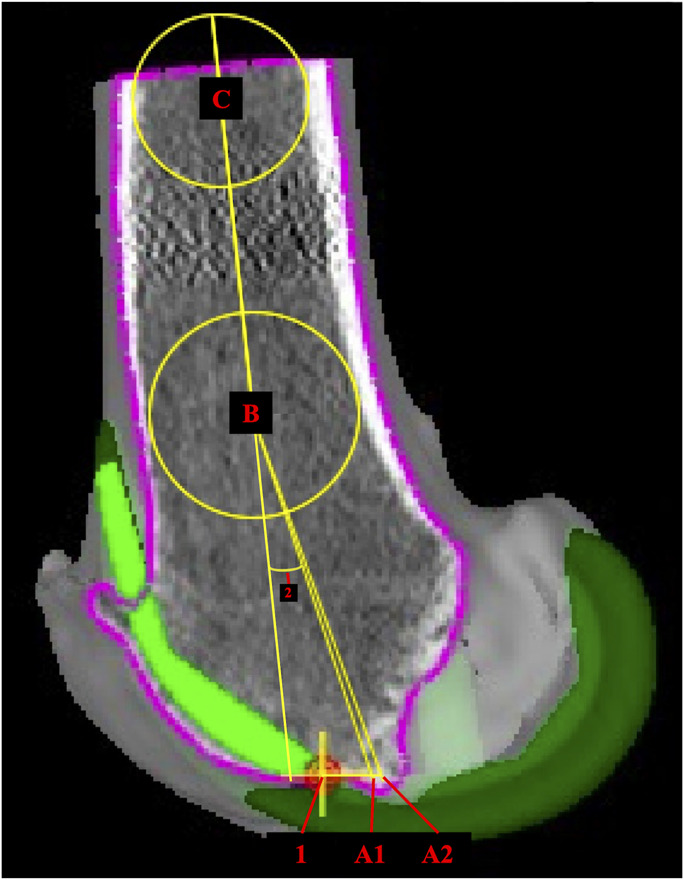
Methods: This is a single-center academic observational study including adult patients undergoing primary RA-TKA by 3 fellowship-trained arthroplasty surgeons between August 2023 and February 2024. Final femoral implant characteristics after knee gap balancing were obtained from intraoperative computerized tomography screenshots. Sagittal angle measurements of rIMN trajectory based on distal femoral nail start point and center of femoral shaft were measured using ImageJ software and compared with several variables with a focus on femoral component flexion for 10-mm and 12-mm nail sizes.
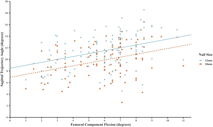
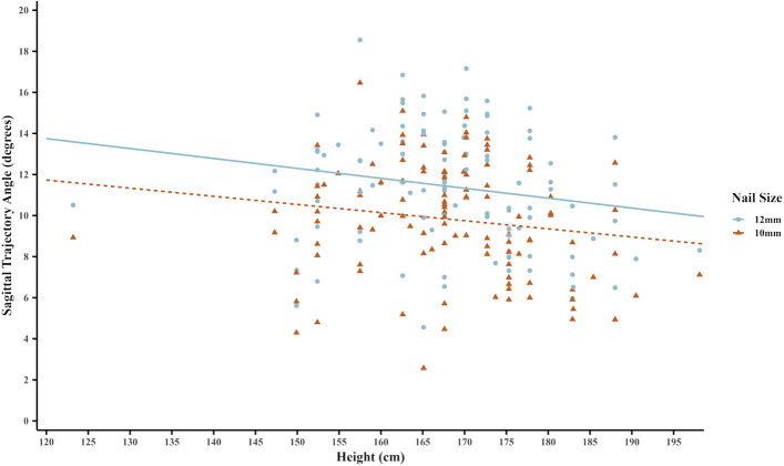
Results: A total of 111 patients (61 female and 50 male) with a mean age of 67 years (SD 10) and height 168 cm (SD 11) were included. The mean femoral component flexion was 5.87° (SD 2.13), and the mean nail sagittal trajectory angle was 9.83° (SD 2.79) and 11.43° (SD 2.88) for the 10-mm and 12-mm nails, respectively. There was a significant linear correlation of a 0.50° increase in the mean sagittal angle for every 1.0° increase in femoral component flexion for both nail sizes (p < 0.001).
Conclusions: There is a linear correlation between femoral component flexion and rIMN sagittal trajectory angle. The mean sagittal angulation was approximately 10°, and the mean component flexion was 6°. As number of RA-TKAs performed nationally is expected to increase, a rIMN with a distal bend designed to compensate for femoral component flexion could be considered to limit hyperextension deformity in distal femur PPFx fixation.
Level of evidence: Level IV. See Instructions for Authors for a complete description of levels of evidence.

 求助内容:
求助内容: 应助结果提醒方式:


