Scarlett Cheong, Rishabh Gupta, Sharada Kadaba Sridhar, Alex J Hall, Michael Frankel, David W Wright, Yuk Y Sham, Uzma Samadani
{"title":"黄体酮在创伤性脑损伤中的应用,实验临床治疗ⅲ:再谈:脑成像分割和生物标志物水平对创伤性脑损伤的客观分类。","authors":"Scarlett Cheong, Rishabh Gupta, Sharada Kadaba Sridhar, Alex J Hall, Michael Frankel, David W Wright, Yuk Y Sham, Uzma Samadani","doi":"10.1097/CCE.0000000000001306","DOIUrl":null,"url":null,"abstract":"<p><strong>Objective: </strong>This post hoc study of the Progesterone for Traumatic Brain Injury, Experimental Clinical Treatment (ProTECT) III trial investigates whether improving traumatic brain injury (TBI) classification, using serum biomarkers (glial fibrillary acidic protein [GFAP] and ubiquitin carboxyl-terminal esterase L1 [UCH-L1]) and algorithmically assessed total lesion volume, could identify a subset of responders to progesterone treatment, beyond broad measures like the Glasgow Coma Scale (GCS) and Glasgow Outcome Scale-Extended (GOS-E), which may fail to capture subtle changes in TBI recovery.</p><p><strong>Design: </strong>Brain lesion volumes on CT scans were quantified using Brain Lesion Analysis and Segmentation Tool for CT. Patients were classified into true-positive and true-negative groups based on an optimization scheme to determine a threshold that maximizes agreement between radiological assessment and objectively measured lesion volume. True-positives were further categorized into low (> 0.2-10 mL), medium (> 10-50 mL), and high (> 50 mL) lesion volumes for analysis with protein biomarkers and injury severity. Correlation analyses linked Rotterdam scores (RSs) with biomarker levels and lesion volumes, whereas Welch's t-test evaluated biomarker differences between groups and progesterone's effects.</p><p><strong>Setting: </strong>Forty-nine level 1 trauma centers in the United States.</p><p><strong>Patient: </strong>Patients with moderate-to-severe TBI.</p><p><strong>Interventions: </strong>Progesterone.</p><p><strong>Measurements and main results: </strong>GFAP and UCH-L1 levels were significantly higher in true-positive cases with low to medium lesion volume. Only UCH-L1 differed between progesterone and placebo groups at 48 hours. Both biomarkers and lesion volume in the true-positive group correlated with the RS. No sex-specific or treatment differences were found.</p><p><strong>Conclusions: </strong>This study reaffirms elevated levels of GFAP and UCH-L1 as biomarkers for detecting TBI in patients with brain lesions and for predicting clinical outcomes. Despite improved classification using CT-imaging segmentation and serum biomarkers, we did not identify a subset of progesterone responders within 24 or 48 hours of progesterone treatment. More rigorous and quantifiable measures for classifying the nature of injury may be needed to enable development of therapeutics as neither serum markers nor algorithmic CT analysis performed better than the older metrics of Rotterdam or GCS metrics.</p>","PeriodicalId":93957,"journal":{"name":"Critical care explorations","volume":"7 9","pages":"e1306"},"PeriodicalIF":2.7000,"publicationDate":"2025-09-05","publicationTypes":"Journal Article","fieldsOfStudy":null,"isOpenAccess":false,"openAccessPdf":"https://www.ncbi.nlm.nih.gov/pmc/articles/PMC12417008/pdf/","citationCount":"0","resultStr":"{\"title\":\"Progesterone for Traumatic Brain Injury, Experimental Clinical Treatment III Trial Revisited: Objective Classification of Traumatic Brain Injury With Brain Imaging Segmentation and Biomarker Levels.\",\"authors\":\"Scarlett Cheong, Rishabh Gupta, Sharada Kadaba Sridhar, Alex J Hall, Michael Frankel, David W Wright, Yuk Y Sham, Uzma Samadani\",\"doi\":\"10.1097/CCE.0000000000001306\",\"DOIUrl\":null,\"url\":null,\"abstract\":\"<p><strong>Objective: </strong>This post hoc study of the Progesterone for Traumatic Brain Injury, Experimental Clinical Treatment (ProTECT) III trial investigates whether improving traumatic brain injury (TBI) classification, using serum biomarkers (glial fibrillary acidic protein [GFAP] and ubiquitin carboxyl-terminal esterase L1 [UCH-L1]) and algorithmically assessed total lesion volume, could identify a subset of responders to progesterone treatment, beyond broad measures like the Glasgow Coma Scale (GCS) and Glasgow Outcome Scale-Extended (GOS-E), which may fail to capture subtle changes in TBI recovery.</p><p><strong>Design: </strong>Brain lesion volumes on CT scans were quantified using Brain Lesion Analysis and Segmentation Tool for CT. Patients were classified into true-positive and true-negative groups based on an optimization scheme to determine a threshold that maximizes agreement between radiological assessment and objectively measured lesion volume. True-positives were further categorized into low (> 0.2-10 mL), medium (> 10-50 mL), and high (> 50 mL) lesion volumes for analysis with protein biomarkers and injury severity. Correlation analyses linked Rotterdam scores (RSs) with biomarker levels and lesion volumes, whereas Welch's t-test evaluated biomarker differences between groups and progesterone's effects.</p><p><strong>Setting: </strong>Forty-nine level 1 trauma centers in the United States.</p><p><strong>Patient: </strong>Patients with moderate-to-severe TBI.</p><p><strong>Interventions: </strong>Progesterone.</p><p><strong>Measurements and main results: </strong>GFAP and UCH-L1 levels were significantly higher in true-positive cases with low to medium lesion volume. Only UCH-L1 differed between progesterone and placebo groups at 48 hours. Both biomarkers and lesion volume in the true-positive group correlated with the RS. No sex-specific or treatment differences were found.</p><p><strong>Conclusions: </strong>This study reaffirms elevated levels of GFAP and UCH-L1 as biomarkers for detecting TBI in patients with brain lesions and for predicting clinical outcomes. Despite improved classification using CT-imaging segmentation and serum biomarkers, we did not identify a subset of progesterone responders within 24 or 48 hours of progesterone treatment. More rigorous and quantifiable measures for classifying the nature of injury may be needed to enable development of therapeutics as neither serum markers nor algorithmic CT analysis performed better than the older metrics of Rotterdam or GCS metrics.</p>\",\"PeriodicalId\":93957,\"journal\":{\"name\":\"Critical care explorations\",\"volume\":\"7 9\",\"pages\":\"e1306\"},\"PeriodicalIF\":2.7000,\"publicationDate\":\"2025-09-05\",\"publicationTypes\":\"Journal Article\",\"fieldsOfStudy\":null,\"isOpenAccess\":false,\"openAccessPdf\":\"https://www.ncbi.nlm.nih.gov/pmc/articles/PMC12417008/pdf/\",\"citationCount\":\"0\",\"resultStr\":null,\"platform\":\"Semanticscholar\",\"paperid\":null,\"PeriodicalName\":\"Critical care explorations\",\"FirstCategoryId\":\"1085\",\"ListUrlMain\":\"https://doi.org/10.1097/CCE.0000000000001306\",\"RegionNum\":0,\"RegionCategory\":null,\"ArticlePicture\":[],\"TitleCN\":null,\"AbstractTextCN\":null,\"PMCID\":null,\"EPubDate\":\"2025/9/1 0:00:00\",\"PubModel\":\"eCollection\",\"JCR\":\"Q4\",\"JCRName\":\"Medicine\",\"Score\":null,\"Total\":0}","platform":"Semanticscholar","paperid":null,"PeriodicalName":"Critical care explorations","FirstCategoryId":"1085","ListUrlMain":"https://doi.org/10.1097/CCE.0000000000001306","RegionNum":0,"RegionCategory":null,"ArticlePicture":[],"TitleCN":null,"AbstractTextCN":null,"PMCID":null,"EPubDate":"2025/9/1 0:00:00","PubModel":"eCollection","JCR":"Q4","JCRName":"Medicine","Score":null,"Total":0}
Progesterone for Traumatic Brain Injury, Experimental Clinical Treatment III Trial Revisited: Objective Classification of Traumatic Brain Injury With Brain Imaging Segmentation and Biomarker Levels.
Objective: This post hoc study of the Progesterone for Traumatic Brain Injury, Experimental Clinical Treatment (ProTECT) III trial investigates whether improving traumatic brain injury (TBI) classification, using serum biomarkers (glial fibrillary acidic protein [GFAP] and ubiquitin carboxyl-terminal esterase L1 [UCH-L1]) and algorithmically assessed total lesion volume, could identify a subset of responders to progesterone treatment, beyond broad measures like the Glasgow Coma Scale (GCS) and Glasgow Outcome Scale-Extended (GOS-E), which may fail to capture subtle changes in TBI recovery.
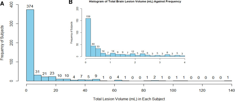
Design: Brain lesion volumes on CT scans were quantified using Brain Lesion Analysis and Segmentation Tool for CT. Patients were classified into true-positive and true-negative groups based on an optimization scheme to determine a threshold that maximizes agreement between radiological assessment and objectively measured lesion volume. True-positives were further categorized into low (> 0.2-10 mL), medium (> 10-50 mL), and high (> 50 mL) lesion volumes for analysis with protein biomarkers and injury severity. Correlation analyses linked Rotterdam scores (RSs) with biomarker levels and lesion volumes, whereas Welch's t-test evaluated biomarker differences between groups and progesterone's effects.
Setting: Forty-nine level 1 trauma centers in the United States.
Patient: Patients with moderate-to-severe TBI.
Interventions: Progesterone.
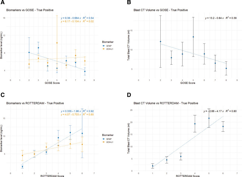
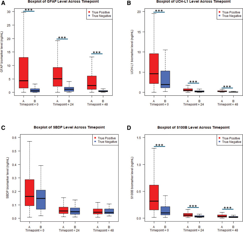
Measurements and main results: GFAP and UCH-L1 levels were significantly higher in true-positive cases with low to medium lesion volume. Only UCH-L1 differed between progesterone and placebo groups at 48 hours. Both biomarkers and lesion volume in the true-positive group correlated with the RS. No sex-specific or treatment differences were found.
Conclusions: This study reaffirms elevated levels of GFAP and UCH-L1 as biomarkers for detecting TBI in patients with brain lesions and for predicting clinical outcomes. Despite improved classification using CT-imaging segmentation and serum biomarkers, we did not identify a subset of progesterone responders within 24 or 48 hours of progesterone treatment. More rigorous and quantifiable measures for classifying the nature of injury may be needed to enable development of therapeutics as neither serum markers nor algorithmic CT analysis performed better than the older metrics of Rotterdam or GCS metrics.

 求助内容:
求助内容: 应助结果提醒方式:


