Luke J. Vano, Robert A. McCutcheon, Jan Sedlacik, Grazia Rutigliano, Stephen J. Kaar, Valeria Finelli, Maria C. Lobo, Alaine Berry, Ben Statton, Amir Fazlollahi, Ian P. Everall, Oliver D. Howes
{"title":"低皮质下铁、白质髓磷脂和少突胶质细胞在精神分裂症中的作用:一项定量易感性映射和弥散张量成像研究","authors":"Luke J. Vano, Robert A. McCutcheon, Jan Sedlacik, Grazia Rutigliano, Stephen J. Kaar, Valeria Finelli, Maria C. Lobo, Alaine Berry, Ben Statton, Amir Fazlollahi, Ian P. Everall, Oliver D. Howes","doi":"10.1038/s41380-025-03195-7","DOIUrl":null,"url":null,"abstract":"<p>Iron—the most abundant magnetic brain substance—is essential for many biological processes, including dopamine and myelin synthesis. Quantitative susceptibility mapping (QSM) MRI has recently linked altered subcortical magnetic susceptibility (χ) to schizophrenia. Since χ is increased by iron and decreased by myelin, abnormal levels of either could underlie these QSM differences. In white matter tracts, magnetic susceptibility anisotropy (δχ) serves as a myelin-specific marker that is insensitive to iron content. To clarify the origin of case-control χ differences, we employed QSM in 85 individuals with schizophrenia, from first-episode mental health teams, and 86 healthy controls. A subset also underwent diffusion tensor imaging (DTI) to calculate subcortical tissue mean diffusivity, which inversely correlates with myelin concentration and fractional anisotropy. White matter δχ was calculated by combining QSM and DTI. Schizophrenia was associated with lower subcortical χ (d = −0.36, p = 0.023). This was significant in the caudate nucleus (d = −0.37, p = 0.037), putamen (d = −0.36, p = 0.037), globus pallidus (d = −0.57, p = 0.001), and SN-VTA (as previously reported). Additionally, schizophrenia was linked to higher subcortical mean diffusivity (d = 0.44, p = 0.018), and lower white matter δχ (d = −0.37, p = 0.047). These findings suggest that both subcortical iron and brain myelin levels are lower in schizophrenia. By comparing our voxelwise χ maps with postmortem gene expression data, we reveal that regions with lower subcortical χ in schizophrenia are enriched for oligodendrocyte-related genes (p < 0.001). As oligodendrocytes are both the most iron-rich brain cells and essential for myelin synthesis, our results implicate oligodendrocyte dysfunction in schizophrenia pathophysiology.</p>","PeriodicalId":19008,"journal":{"name":"Molecular Psychiatry","volume":"48 1","pages":""},"PeriodicalIF":10.1000,"publicationDate":"2025-09-05","publicationTypes":"Journal Article","fieldsOfStudy":null,"isOpenAccess":false,"openAccessPdf":"","citationCount":"0","resultStr":"{\"title\":\"The role of low subcortical iron, white matter myelin, and oligodendrocytes in schizophrenia: a quantitative susceptibility mapping and diffusion tensor imaging study\",\"authors\":\"Luke J. Vano, Robert A. McCutcheon, Jan Sedlacik, Grazia Rutigliano, Stephen J. Kaar, Valeria Finelli, Maria C. Lobo, Alaine Berry, Ben Statton, Amir Fazlollahi, Ian P. Everall, Oliver D. Howes\",\"doi\":\"10.1038/s41380-025-03195-7\",\"DOIUrl\":null,\"url\":null,\"abstract\":\"<p>Iron—the most abundant magnetic brain substance—is essential for many biological processes, including dopamine and myelin synthesis. Quantitative susceptibility mapping (QSM) MRI has recently linked altered subcortical magnetic susceptibility (χ) to schizophrenia. Since χ is increased by iron and decreased by myelin, abnormal levels of either could underlie these QSM differences. In white matter tracts, magnetic susceptibility anisotropy (δχ) serves as a myelin-specific marker that is insensitive to iron content. To clarify the origin of case-control χ differences, we employed QSM in 85 individuals with schizophrenia, from first-episode mental health teams, and 86 healthy controls. A subset also underwent diffusion tensor imaging (DTI) to calculate subcortical tissue mean diffusivity, which inversely correlates with myelin concentration and fractional anisotropy. White matter δχ was calculated by combining QSM and DTI. Schizophrenia was associated with lower subcortical χ (d = −0.36, p = 0.023). This was significant in the caudate nucleus (d = −0.37, p = 0.037), putamen (d = −0.36, p = 0.037), globus pallidus (d = −0.57, p = 0.001), and SN-VTA (as previously reported). Additionally, schizophrenia was linked to higher subcortical mean diffusivity (d = 0.44, p = 0.018), and lower white matter δχ (d = −0.37, p = 0.047). These findings suggest that both subcortical iron and brain myelin levels are lower in schizophrenia. By comparing our voxelwise χ maps with postmortem gene expression data, we reveal that regions with lower subcortical χ in schizophrenia are enriched for oligodendrocyte-related genes (p < 0.001). As oligodendrocytes are both the most iron-rich brain cells and essential for myelin synthesis, our results implicate oligodendrocyte dysfunction in schizophrenia pathophysiology.</p>\",\"PeriodicalId\":19008,\"journal\":{\"name\":\"Molecular Psychiatry\",\"volume\":\"48 1\",\"pages\":\"\"},\"PeriodicalIF\":10.1000,\"publicationDate\":\"2025-09-05\",\"publicationTypes\":\"Journal Article\",\"fieldsOfStudy\":null,\"isOpenAccess\":false,\"openAccessPdf\":\"\",\"citationCount\":\"0\",\"resultStr\":null,\"platform\":\"Semanticscholar\",\"paperid\":null,\"PeriodicalName\":\"Molecular Psychiatry\",\"FirstCategoryId\":\"3\",\"ListUrlMain\":\"https://doi.org/10.1038/s41380-025-03195-7\",\"RegionNum\":1,\"RegionCategory\":\"医学\",\"ArticlePicture\":[],\"TitleCN\":null,\"AbstractTextCN\":null,\"PMCID\":null,\"EPubDate\":\"\",\"PubModel\":\"\",\"JCR\":\"Q1\",\"JCRName\":\"BIOCHEMISTRY & MOLECULAR BIOLOGY\",\"Score\":null,\"Total\":0}","platform":"Semanticscholar","paperid":null,"PeriodicalName":"Molecular Psychiatry","FirstCategoryId":"3","ListUrlMain":"https://doi.org/10.1038/s41380-025-03195-7","RegionNum":1,"RegionCategory":"医学","ArticlePicture":[],"TitleCN":null,"AbstractTextCN":null,"PMCID":null,"EPubDate":"","PubModel":"","JCR":"Q1","JCRName":"BIOCHEMISTRY & MOLECULAR BIOLOGY","Score":null,"Total":0}
引用次数: 0
摘要
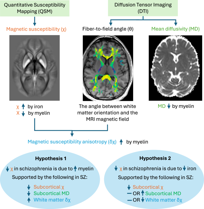
铁是大脑中最丰富的磁性物质,对许多生物过程至关重要,包括多巴胺和髓磷脂的合成。定量易感性制图(QSM) MRI最近将皮质下磁化率(χ)的改变与精神分裂症联系起来。由于χ因铁升高而髓磷脂降低,因此异常水平可能是这些QSM差异的基础。在白质束中,磁化率各向异性(δχ)作为髓磷脂特异性标记,对铁含量不敏感。为了澄清病例-对照χ差异的来源,我们对来自首发精神卫生团队的85名精神分裂症患者和86名健康对照者采用了QSM。一个子集还接受了扩散张量成像(DTI)来计算皮层下组织的平均扩散率,这与髓磷脂浓度和分数各向异性呈负相关。结合QSM和DTI计算白质δχ。精神分裂症与较低的皮质下χ (d = - 0.36, p = 0.023)相关。这在尾状核(d = - 0.37, p = 0.037)、壳核(d = - 0.36, p = 0.037)、苍白球(d = - 0.57, p = 0.001)和SN-VTA(如先前报道)中具有显著意义。此外,精神分裂症与较高的皮层下平均扩散率(d = 0.44, p = 0.018)和较低的白质δχ (d = - 0.37, p = 0.047)有关。这些发现表明,精神分裂症患者的皮质下铁和脑髓磷脂水平都较低。通过将我们的体向χ图与死后基因表达数据进行比较,我们发现精神分裂症患者皮质下χ较低的区域富含少突胶质细胞相关基因(p < 0.001)。由于少突胶质细胞是最富铁的脑细胞,也是髓磷脂合成所必需的,因此我们的研究结果暗示少突胶质细胞功能障碍与精神分裂症的病理生理有关。
The role of low subcortical iron, white matter myelin, and oligodendrocytes in schizophrenia: a quantitative susceptibility mapping and diffusion tensor imaging study
Iron—the most abundant magnetic brain substance—is essential for many biological processes, including dopamine and myelin synthesis. Quantitative susceptibility mapping (QSM) MRI has recently linked altered subcortical magnetic susceptibility (χ) to schizophrenia. Since χ is increased by iron and decreased by myelin, abnormal levels of either could underlie these QSM differences. In white matter tracts, magnetic susceptibility anisotropy (δχ) serves as a myelin-specific marker that is insensitive to iron content. To clarify the origin of case-control χ differences, we employed QSM in 85 individuals with schizophrenia, from first-episode mental health teams, and 86 healthy controls. A subset also underwent diffusion tensor imaging (DTI) to calculate subcortical tissue mean diffusivity, which inversely correlates with myelin concentration and fractional anisotropy. White matter δχ was calculated by combining QSM and DTI. Schizophrenia was associated with lower subcortical χ (d = −0.36, p = 0.023). This was significant in the caudate nucleus (d = −0.37, p = 0.037), putamen (d = −0.36, p = 0.037), globus pallidus (d = −0.57, p = 0.001), and SN-VTA (as previously reported). Additionally, schizophrenia was linked to higher subcortical mean diffusivity (d = 0.44, p = 0.018), and lower white matter δχ (d = −0.37, p = 0.047). These findings suggest that both subcortical iron and brain myelin levels are lower in schizophrenia. By comparing our voxelwise χ maps with postmortem gene expression data, we reveal that regions with lower subcortical χ in schizophrenia are enriched for oligodendrocyte-related genes (p < 0.001). As oligodendrocytes are both the most iron-rich brain cells and essential for myelin synthesis, our results implicate oligodendrocyte dysfunction in schizophrenia pathophysiology.
期刊介绍:
Molecular Psychiatry focuses on publishing research that aims to uncover the biological mechanisms behind psychiatric disorders and their treatment. The journal emphasizes studies that bridge pre-clinical and clinical research, covering cellular, molecular, integrative, clinical, imaging, and psychopharmacology levels.
 求助内容:
求助内容: 应助结果提醒方式:


