白介素-6调节急性穷竭运动后肾脏内质网应激信号和线粒体蛋白复合物。
IF 3.2
3区 生物学
Q3 CELL BIOLOGY
引用次数: 0
摘要
内质网(ER)的稳态受到一个被称为未折叠蛋白反应(UPR)的自适应信号网络的密切调节,该信号网络与炎症途径密切相关。然而,体育锻炼会增加血浆中白细胞介素-6 (IL-6)的浓度,IL-6具有促炎和抗炎特性,介导内质网功能和线粒体代谢,这使得其研究与生理和病理背景相关。在肾脏疾病中,IL-6水平可有效预测死亡风险。为了阐明运动诱导的IL-6升高、内质网应激和肾脏生理之间的关系,我们探讨了急性穷尽性运动对IL-6敲除(KO)小鼠肾脏内质网应激相关蛋白和线粒体呼吸链靶点的影响。WT和IL-6 KO小鼠根据每种表型分为两个亚组:久坐(Sed)和1小时(急性运动后1小时;Ex-1h)。取出肾脏,准备进行组织学、逆转录定量聚合酶链反应(RT-qPCR)和免疫印迹分析。总之,IL-6 KO小鼠肾脏中脱颗粒肥大细胞较低。IL-6 KO小鼠表现出运动能力下降。在WT组和KO组中,Hspa5 mRNA水平在剧烈运动后显著升高,但Il-10仅在KO组中升高。此外,运动后IL-6 KO小鼠的Ddit3表达显著降低,表明没有IL-6时内质网应激反应减弱。在蛋白水平上,运动后IL-6 KO小鼠的ATF6α表达显著升高。关于线粒体蛋白复合物,我们观察到WT Ex-1h组的线粒体复合物IV和CII蛋白水平低于WT Sed组。同时,IL-6的缺乏似乎并没有改变大多数线粒体复合物在急性运动后的表达。此外,公开可用的人类基因表达数据集支持我们的发现,表明IL-6信号和热休克蛋白(HSPs)上调,同时降低急性穷尽运动后人类白细胞中线粒体呼吸复合体mRNA水平。研究结果表明,IL-6可能调节急性运动后肾脏内质网应激和细胞因子反应的特定成分。本文章由计算机程序翻译,如有差异,请以英文原文为准。
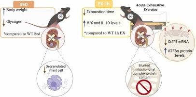
Interleukin-6 modulates endoplasmic reticulum stress signaling and mitochondrial protein complexes in the kidney following acute exhaustive exercise
Endoplasmic Reticulum (ER) homeostasis is closely regulated by an adaptive signaling network identified as the unfolded protein response (UPR), which is tightly related to the inflammatory pathway. However, physical exercise increases plasma concentrations of interleukin-6 (IL-6), which exhibits both pro- and anti-inflammatory properties that mediate ER function and mitochondrial metabolism, making its investigation relevant in physiological and pathological contexts. In kidney diseases, the IL-6 levels are effective in predicting mortality risk. To elucidate the relationship between exercise-induced IL-6 elevation, ER stress, and renal physiology, we explored the impact of an acute exhaustive exercise on the ER stress-related proteins and mitochondrial respiratory chain targets in the kidneys of IL-6 knockout (KO) mice. WT and IL-6 KO mice were divided into two subgroups for each phenotype: sedentary (Sed) and 1 h (after 1 h of acute exercise; Ex-1h). The kidneys were removed and prepared for histological, reverse transcription-quantitative polymerase chain reaction (RT-qPCR), and immunoblotting analysis. In summary, IL-6 KO mice had lower degranulated mast cells in the kidney. IL-6 KO mice exhibited reduced exercise performance. The Hspa5 mRNA levels were significantly increased in response to acute exhaustive exercise in both WT and KO groups, but Il-10 increased only in response to exercise in the KO group. Additionally, Ddit3 expression was significantly lower in IL-6 KO mice post-exercise, suggesting a blunted ER stress response without IL-6. At the protein levels, ATF6α expression was notably elevated in IL-6 KO mice following exercise. Regarding mitochondrial protein complexes, we observed lower protein levels of mitochondrial complex IV and CII in the WT Ex-1h group than in the WT Sed. At the same time, the absence of IL-6 did not seem to modify the expression of most mitochondrial complexes in response to acute exercise. Also, publicly available gene expression datasets in humans support our findings, indicating the upregulation of IL-6 signaling and heat shock proteins (HSPs), while decreasing mitochondrial respiratory complex mRNA levels in white blood cells of humans following acute exhaustive exercise. The findings indicate that IL-6 may modulate specific components of ER stress and cytokine responses in the kidney after acute exercise.
求助全文
通过发布文献求助,成功后即可免费获取论文全文。
去求助
来源期刊
Cell Stress & Chaperones
生物-细胞生物学
CiteScore
7.60
自引率
2.60%
发文量
59
审稿时长
6-12 weeks
期刊介绍:
Cell Stress and Chaperones is an integrative journal that bridges the gap between laboratory model systems and natural populations. The journal captures the eclectic spirit of the cellular stress response field in a single, concentrated source of current information. Major emphasis is placed on the effects of climate change on individual species in the natural environment and their capacity to adapt. This emphasis expands our focus on stress biology and medicine by linking climate change effects to research on cellular stress responses of animals, micro-organisms and plants.
 求助内容:
求助内容: 应助结果提醒方式:


