Klevis Mihali, Birgit Markus, Bernhard Schieffer, Marcus Bauer, Julian Kreutz
{"title":"跳出框框思考:罕见的四尖瓣主动脉瓣作为心力衰竭和心房颤动的未被充分认识的原因的病例报告。","authors":"Klevis Mihali, Birgit Markus, Bernhard Schieffer, Marcus Bauer, Julian Kreutz","doi":"10.14740/jmc5153","DOIUrl":null,"url":null,"abstract":"<p><p>Quadricuspid aortic valve (QAV) is a rare congenital anomaly with an estimated incidence of 0.008% to 0.043% based on autopsy and echocardiographic studies. Although often asymptomatic, it can lead to progressive aortic regurgitation (AR), left ventricular (LV) dysfunction, and arrhythmias such as atrial fibrillation (AF). Due to its rarity, QAV is often misdiagnosed or discovered incidentally, highlighting the need for advanced cardiac imaging in young patients presenting with unexplained heart failure symptoms and arrhythmias. We present the case of a 41-year-old female patient who was admitted with new-onset dyspnea classified as New York Heart Association (NYHA) class III and palpitations due to persistent AF with a European Heart Rhythm Association (EHRA) symptom class 2b. There was no family history of congenital or structural heart disease, with arterial hypertension being the only identified predisposing condition. Initial transthoracic echocardiography revealed moderate AR, but more detailed transesophageal echocardiography performed before pulmonary vein isolation incidentally revealed a QAV. Further cardiac magnetic resonance imaging confirmed normal aortic root dimensions with early LV remodeling. The patient was managed conservatively with rate control, anticoagulation, and regular follow-up to monitor disease progression. This case highlights the importance of advanced imaging techniques in the diagnosis of rare structural heart abnormalities in young patients presenting with unexplained heart failure symptoms and arrhythmias. Early identification of QAV allows for timely medical intervention, optimal patient monitoring, and prevention of long-term complications. Regular follow-up is essential to monitor disease progression and determine the need for surgical intervention.</p>","PeriodicalId":101328,"journal":{"name":"Journal of medical cases","volume":"16 8","pages":"277-281"},"PeriodicalIF":0.9000,"publicationDate":"2025-08-07","publicationTypes":"Journal Article","fieldsOfStudy":null,"isOpenAccess":false,"openAccessPdf":"https://www.ncbi.nlm.nih.gov/pmc/articles/PMC12404117/pdf/","citationCount":"0","resultStr":"{\"title\":\"Thinking Outside the Box: Case Report of a Rare Quadricuspid Aortic Valve as an Underrecognized Cause of Heart Failure and Atrial Fibrillation.\",\"authors\":\"Klevis Mihali, Birgit Markus, Bernhard Schieffer, Marcus Bauer, Julian Kreutz\",\"doi\":\"10.14740/jmc5153\",\"DOIUrl\":null,\"url\":null,\"abstract\":\"<p><p>Quadricuspid aortic valve (QAV) is a rare congenital anomaly with an estimated incidence of 0.008% to 0.043% based on autopsy and echocardiographic studies. Although often asymptomatic, it can lead to progressive aortic regurgitation (AR), left ventricular (LV) dysfunction, and arrhythmias such as atrial fibrillation (AF). Due to its rarity, QAV is often misdiagnosed or discovered incidentally, highlighting the need for advanced cardiac imaging in young patients presenting with unexplained heart failure symptoms and arrhythmias. We present the case of a 41-year-old female patient who was admitted with new-onset dyspnea classified as New York Heart Association (NYHA) class III and palpitations due to persistent AF with a European Heart Rhythm Association (EHRA) symptom class 2b. There was no family history of congenital or structural heart disease, with arterial hypertension being the only identified predisposing condition. Initial transthoracic echocardiography revealed moderate AR, but more detailed transesophageal echocardiography performed before pulmonary vein isolation incidentally revealed a QAV. Further cardiac magnetic resonance imaging confirmed normal aortic root dimensions with early LV remodeling. The patient was managed conservatively with rate control, anticoagulation, and regular follow-up to monitor disease progression. This case highlights the importance of advanced imaging techniques in the diagnosis of rare structural heart abnormalities in young patients presenting with unexplained heart failure symptoms and arrhythmias. Early identification of QAV allows for timely medical intervention, optimal patient monitoring, and prevention of long-term complications. Regular follow-up is essential to monitor disease progression and determine the need for surgical intervention.</p>\",\"PeriodicalId\":101328,\"journal\":{\"name\":\"Journal of medical cases\",\"volume\":\"16 8\",\"pages\":\"277-281\"},\"PeriodicalIF\":0.9000,\"publicationDate\":\"2025-08-07\",\"publicationTypes\":\"Journal Article\",\"fieldsOfStudy\":null,\"isOpenAccess\":false,\"openAccessPdf\":\"https://www.ncbi.nlm.nih.gov/pmc/articles/PMC12404117/pdf/\",\"citationCount\":\"0\",\"resultStr\":null,\"platform\":\"Semanticscholar\",\"paperid\":null,\"PeriodicalName\":\"Journal of medical cases\",\"FirstCategoryId\":\"1085\",\"ListUrlMain\":\"https://doi.org/10.14740/jmc5153\",\"RegionNum\":0,\"RegionCategory\":null,\"ArticlePicture\":[],\"TitleCN\":null,\"AbstractTextCN\":null,\"PMCID\":null,\"EPubDate\":\"2025/8/1 0:00:00\",\"PubModel\":\"eCollection\",\"JCR\":\"\",\"JCRName\":\"\",\"Score\":null,\"Total\":0}","platform":"Semanticscholar","paperid":null,"PeriodicalName":"Journal of medical cases","FirstCategoryId":"1085","ListUrlMain":"https://doi.org/10.14740/jmc5153","RegionNum":0,"RegionCategory":null,"ArticlePicture":[],"TitleCN":null,"AbstractTextCN":null,"PMCID":null,"EPubDate":"2025/8/1 0:00:00","PubModel":"eCollection","JCR":"","JCRName":"","Score":null,"Total":0}
Thinking Outside the Box: Case Report of a Rare Quadricuspid Aortic Valve as an Underrecognized Cause of Heart Failure and Atrial Fibrillation.
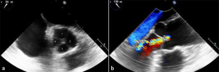
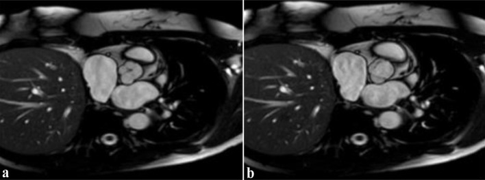
Quadricuspid aortic valve (QAV) is a rare congenital anomaly with an estimated incidence of 0.008% to 0.043% based on autopsy and echocardiographic studies. Although often asymptomatic, it can lead to progressive aortic regurgitation (AR), left ventricular (LV) dysfunction, and arrhythmias such as atrial fibrillation (AF). Due to its rarity, QAV is often misdiagnosed or discovered incidentally, highlighting the need for advanced cardiac imaging in young patients presenting with unexplained heart failure symptoms and arrhythmias. We present the case of a 41-year-old female patient who was admitted with new-onset dyspnea classified as New York Heart Association (NYHA) class III and palpitations due to persistent AF with a European Heart Rhythm Association (EHRA) symptom class 2b. There was no family history of congenital or structural heart disease, with arterial hypertension being the only identified predisposing condition. Initial transthoracic echocardiography revealed moderate AR, but more detailed transesophageal echocardiography performed before pulmonary vein isolation incidentally revealed a QAV. Further cardiac magnetic resonance imaging confirmed normal aortic root dimensions with early LV remodeling. The patient was managed conservatively with rate control, anticoagulation, and regular follow-up to monitor disease progression. This case highlights the importance of advanced imaging techniques in the diagnosis of rare structural heart abnormalities in young patients presenting with unexplained heart failure symptoms and arrhythmias. Early identification of QAV allows for timely medical intervention, optimal patient monitoring, and prevention of long-term complications. Regular follow-up is essential to monitor disease progression and determine the need for surgical intervention.

 求助内容:
求助内容: 应助结果提醒方式:


