Raphael B Takyi, Jeanette Plantin, Sylvain Charron, Marc A Maier, Jean-Claude Baron, Guillaume Turc, Charlotte Rosso, Clément Debacker, Påvel G Lindberg
{"title":"脑卒中后纵向脑老化:神经退行性变的标志及其与上肢运动结果的相关性。","authors":"Raphael B Takyi, Jeanette Plantin, Sylvain Charron, Marc A Maier, Jean-Claude Baron, Guillaume Turc, Charlotte Rosso, Clément Debacker, Påvel G Lindberg","doi":"10.1093/braincomms/fcaf299","DOIUrl":null,"url":null,"abstract":"<p><p>Brain age, as distinct from chronological age, may reveal post-stroke recovery mechanisms, but longitudinal studies tracking brain age are lacking. We explored longitudinal change of brain age post-stroke and its relation to upper limb sensorimotor outcome. T<sub>1</sub>-weighted MRI at baseline (∼3 weeks) and follow-up (3-7 months) post-stroke was used to estimate brain age. Difference to chronological age was calculated as brain age gap (BAG). Grey and white matter changes and lesion location related to increased brain ageing were investigated, controlling for lesion volume. Association between BAG change and upper limb sensorimotor outcome was studied using linear mixed effects regression. Totally, 114 stroke patients with arm/hand hemiparesis were pooled from three studies. BAG significantly increased from baseline to follow-up, a period of ∼6 months, by a mean of 3.62 years (<i>t</i> = -7.31; <i>P</i> < 0.001). Voxel-based morphometry showed that high BAG change was related to reduced grey and white matter volume ipsilesionally, extending beyond the stroke lesion. Voxel-based lesion symptom mapping showed that lesion to thalamocortical projections, internal capsule and corona radiata related to accelerated brain ageing. BAG change was significantly associated with motor outcomes in the sub-acute to chronic phase, as expressed by Fugl-Meyer assessment (<i>β</i> = -5.62, SE = 2.81, <i>t</i> = -2.00, <i>P</i> = 0.05), maximum grip strength (<i>β</i> = -0.14, SE = 0.04, <i>t</i> = -3.36, <i>P</i> = 0.001) and dexterity assessment (<i>β</i> = -0.09, SE = 0.04, <i>t</i> = -2.17, <i>P</i> = 0.03). We demonstrate increased brain ageing within the first few months post-stroke. This secondary neurodegeneration was negatively related to motor outcome. Brain age may be a valid whole-brain probe of individual secondary post-stroke degeneration, relevant for predicting recovery and identifying targets of neural plasticity.</p>","PeriodicalId":93915,"journal":{"name":"Brain communications","volume":"7 5","pages":"fcaf299"},"PeriodicalIF":4.5000,"publicationDate":"2025-08-14","publicationTypes":"Journal Article","fieldsOfStudy":null,"isOpenAccess":false,"openAccessPdf":"https://www.ncbi.nlm.nih.gov/pmc/articles/PMC12399367/pdf/","citationCount":"0","resultStr":"{\"title\":\"Longitudinal brain ageing after stroke: a marker for neurodegeneration and its relevance for upper limb motor outcome.\",\"authors\":\"Raphael B Takyi, Jeanette Plantin, Sylvain Charron, Marc A Maier, Jean-Claude Baron, Guillaume Turc, Charlotte Rosso, Clément Debacker, Påvel G Lindberg\",\"doi\":\"10.1093/braincomms/fcaf299\",\"DOIUrl\":null,\"url\":null,\"abstract\":\"<p><p>Brain age, as distinct from chronological age, may reveal post-stroke recovery mechanisms, but longitudinal studies tracking brain age are lacking. We explored longitudinal change of brain age post-stroke and its relation to upper limb sensorimotor outcome. T<sub>1</sub>-weighted MRI at baseline (∼3 weeks) and follow-up (3-7 months) post-stroke was used to estimate brain age. Difference to chronological age was calculated as brain age gap (BAG). Grey and white matter changes and lesion location related to increased brain ageing were investigated, controlling for lesion volume. Association between BAG change and upper limb sensorimotor outcome was studied using linear mixed effects regression. Totally, 114 stroke patients with arm/hand hemiparesis were pooled from three studies. BAG significantly increased from baseline to follow-up, a period of ∼6 months, by a mean of 3.62 years (<i>t</i> = -7.31; <i>P</i> < 0.001). Voxel-based morphometry showed that high BAG change was related to reduced grey and white matter volume ipsilesionally, extending beyond the stroke lesion. Voxel-based lesion symptom mapping showed that lesion to thalamocortical projections, internal capsule and corona radiata related to accelerated brain ageing. BAG change was significantly associated with motor outcomes in the sub-acute to chronic phase, as expressed by Fugl-Meyer assessment (<i>β</i> = -5.62, SE = 2.81, <i>t</i> = -2.00, <i>P</i> = 0.05), maximum grip strength (<i>β</i> = -0.14, SE = 0.04, <i>t</i> = -3.36, <i>P</i> = 0.001) and dexterity assessment (<i>β</i> = -0.09, SE = 0.04, <i>t</i> = -2.17, <i>P</i> = 0.03). We demonstrate increased brain ageing within the first few months post-stroke. This secondary neurodegeneration was negatively related to motor outcome. Brain age may be a valid whole-brain probe of individual secondary post-stroke degeneration, relevant for predicting recovery and identifying targets of neural plasticity.</p>\",\"PeriodicalId\":93915,\"journal\":{\"name\":\"Brain communications\",\"volume\":\"7 5\",\"pages\":\"fcaf299\"},\"PeriodicalIF\":4.5000,\"publicationDate\":\"2025-08-14\",\"publicationTypes\":\"Journal Article\",\"fieldsOfStudy\":null,\"isOpenAccess\":false,\"openAccessPdf\":\"https://www.ncbi.nlm.nih.gov/pmc/articles/PMC12399367/pdf/\",\"citationCount\":\"0\",\"resultStr\":null,\"platform\":\"Semanticscholar\",\"paperid\":null,\"PeriodicalName\":\"Brain communications\",\"FirstCategoryId\":\"1085\",\"ListUrlMain\":\"https://doi.org/10.1093/braincomms/fcaf299\",\"RegionNum\":0,\"RegionCategory\":null,\"ArticlePicture\":[],\"TitleCN\":null,\"AbstractTextCN\":null,\"PMCID\":null,\"EPubDate\":\"2025/1/1 0:00:00\",\"PubModel\":\"eCollection\",\"JCR\":\"Q1\",\"JCRName\":\"CLINICAL NEUROLOGY\",\"Score\":null,\"Total\":0}","platform":"Semanticscholar","paperid":null,"PeriodicalName":"Brain communications","FirstCategoryId":"1085","ListUrlMain":"https://doi.org/10.1093/braincomms/fcaf299","RegionNum":0,"RegionCategory":null,"ArticlePicture":[],"TitleCN":null,"AbstractTextCN":null,"PMCID":null,"EPubDate":"2025/1/1 0:00:00","PubModel":"eCollection","JCR":"Q1","JCRName":"CLINICAL NEUROLOGY","Score":null,"Total":0}
引用次数: 0
摘要
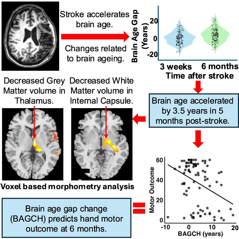
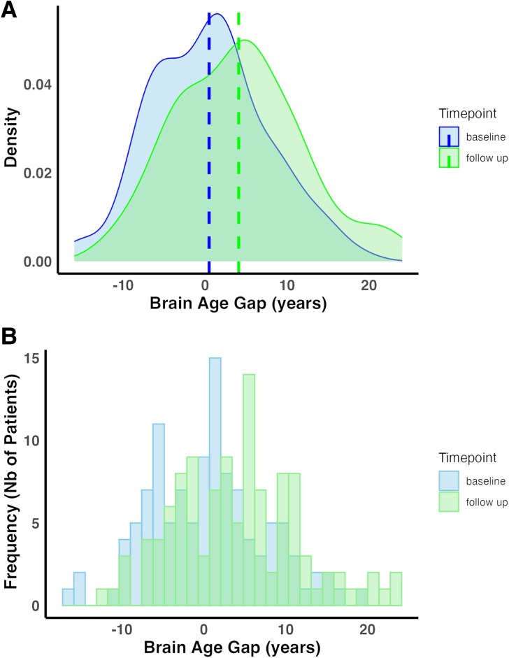
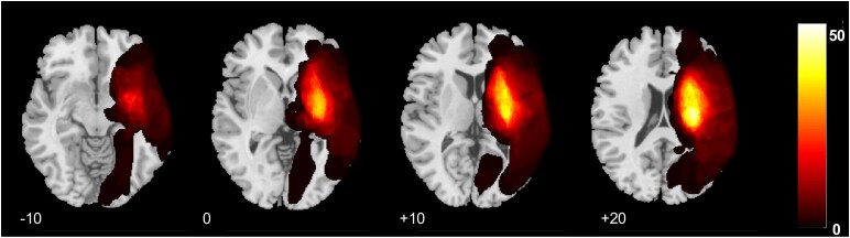
脑年龄不同于实足年龄,可能揭示中风后的恢复机制,但缺乏追踪脑年龄的纵向研究。我们探讨脑卒中后脑年龄的纵向变化及其与上肢感觉运动预后的关系。脑卒中后基线(~ 3周)和随访(3-7个月)的t1加权MRI用于估计脑年龄。与实足年龄的差异计算为脑年龄差距(BAG)。在控制病变体积的情况下,研究了与脑老化增加相关的灰质和白质变化和病变位置。采用线性混合效应回归研究BAG变化与上肢感觉运动预后的关系。共纳入3项研究的114例卒中伴臂/手偏瘫患者。从基线到随访(约6个月),BAG显著增加,平均为3.62年(t = -7.31; P < 0.001)。基于体素的形态测量显示,BAG的高变化与脑灰质和白质体积的减少有关,并延伸到脑卒中病变之外。基于体素的病变症状图谱显示,丘脑皮质突起、内囊和辐射冠与脑加速老化有关。通过fugel - meyer评估(β = -5.62, SE = 2.81, t = -2.00, P = 0.05)、最大握力(β = -0.14, SE = 0.04, t = -3.36, P = 0.001)和灵巧度评估(β = -0.09, SE = 0.04, t = -2.17, P = 0.03), BAG变化与亚急性至慢性期运动结果显著相关。我们证明中风后的头几个月大脑老化加剧。继发性神经变性与运动预后呈负相关。脑年龄可能是个体继发性脑卒中后变性的有效全脑探测,与预测恢复和识别神经可塑性目标相关。
Longitudinal brain ageing after stroke: a marker for neurodegeneration and its relevance for upper limb motor outcome.
Brain age, as distinct from chronological age, may reveal post-stroke recovery mechanisms, but longitudinal studies tracking brain age are lacking. We explored longitudinal change of brain age post-stroke and its relation to upper limb sensorimotor outcome. T1-weighted MRI at baseline (∼3 weeks) and follow-up (3-7 months) post-stroke was used to estimate brain age. Difference to chronological age was calculated as brain age gap (BAG). Grey and white matter changes and lesion location related to increased brain ageing were investigated, controlling for lesion volume. Association between BAG change and upper limb sensorimotor outcome was studied using linear mixed effects regression. Totally, 114 stroke patients with arm/hand hemiparesis were pooled from three studies. BAG significantly increased from baseline to follow-up, a period of ∼6 months, by a mean of 3.62 years (t = -7.31; P < 0.001). Voxel-based morphometry showed that high BAG change was related to reduced grey and white matter volume ipsilesionally, extending beyond the stroke lesion. Voxel-based lesion symptom mapping showed that lesion to thalamocortical projections, internal capsule and corona radiata related to accelerated brain ageing. BAG change was significantly associated with motor outcomes in the sub-acute to chronic phase, as expressed by Fugl-Meyer assessment (β = -5.62, SE = 2.81, t = -2.00, P = 0.05), maximum grip strength (β = -0.14, SE = 0.04, t = -3.36, P = 0.001) and dexterity assessment (β = -0.09, SE = 0.04, t = -2.17, P = 0.03). We demonstrate increased brain ageing within the first few months post-stroke. This secondary neurodegeneration was negatively related to motor outcome. Brain age may be a valid whole-brain probe of individual secondary post-stroke degeneration, relevant for predicting recovery and identifying targets of neural plasticity.

 求助内容:
求助内容: 应助结果提醒方式:


