{"title":"外周嗜碱性粒细胞活化:原发性胆道胆管炎免疫发病机制的隐藏参与者。","authors":"Huan-Qin Han, Jia-Min Bao, Wei Deng, Wei-Fang Guo, Yi-Fan Li, Wei-Qiang Zheng, Hua-Feng Liu","doi":"10.4254/wjh.v17.i8.109685","DOIUrl":null,"url":null,"abstract":"<p><strong>Background: </strong>T helper 17 (Th17) cell infiltration and interleukin (IL)-17 secretion in intrahepatic small bile ducts is a critical driver of immune-mediated injury in primary biliary cholangitis (PBC). IL-6 is an essential upstream activator of Th17 cells. Basophil-derived IL-6 promotes the differentiation of CD4+ T cells and Th1 cells into Th17 cells, thereby regulating their immunological functions.</p><p><strong>Aim: </strong>To investigate the activation status and cytokine expression of basophils in PBC, elucidating potential mechanisms through which basophils contribute to its pathogenesis.</p><p><strong>Methods: </strong>This single-center retrospective case-control study conducted at Guangdong Medical University Affiliated Hospital (China) between September 2019 and August 2024 enrolled 65 consecutive treatment-naïve patients with PBC (PBC group), 65 age- and sex-matched patients with chronic hepatitis B (CHB group), and 65 healthy controls (Normal group). Fourteen participants per group (subgroup) were randomly selected for flow cytometry analysis of basophil proportion, activation markers (CD203c and CD62 L mean fluorescence intensity), IL-6-positive basophils (IL-6+ basophils as a percentage of total basophils), and IL-17-positive T lymphocytes (CD3+CD4+IL-17+ cells) proportion among T cells. Data were analyzed using Kruskal-Wallis and <i>χ</i> <sup>2</sup> tests as appropriate.</p><p><strong>Results: </strong>Routine blood tests revealed significantly higher basophil counts and proportions in the PBC group compared to the CHB and Normal groups (<i>P</i> < 0.001 for both comparisons), with no significant differences between the CHB and Normal groups (<i>P</i> = 0.201). Flow cytometry revealed a higher basophil proportion in the PBC subgroup compared to the CHB (<i>P</i> = 0.011) and Normal subgroups (<i>P</i> < 0.001). The mean fluorescence intensity of CD203c on basophil surfaces was elevated in the PBC subgroup compared to the CHB (<i>P</i> = 0.032) and Normal subgroups (<i>P</i> = 0.039). The proportion of IL-6+ basophils was significantly higher in the PBC subgroup than in the CHB (<i>P</i> < 0.01) and Normal subgroups (<i>P</i> < 0.001). Similarly, the Th17 cell proportion was markedly elevated in the PBC compared to the CHB (<i>P</i> < 0.001) and Normal subgroups (<i>P</i> < 0.001).</p><p><strong>Conclusion: </strong>Patients with PBC have increased peripheral basophil counts with enhanced activation. Activated basophils have increased IL-6 expression, which may indirectly induce Th17 cell proliferation and contribute to PBC pathogenesis.</p>","PeriodicalId":23687,"journal":{"name":"World Journal of Hepatology","volume":"17 8","pages":"109685"},"PeriodicalIF":2.5000,"publicationDate":"2025-08-27","publicationTypes":"Journal Article","fieldsOfStudy":null,"isOpenAccess":false,"openAccessPdf":"https://www.ncbi.nlm.nih.gov/pmc/articles/PMC12400441/pdf/","citationCount":"0","resultStr":"{\"title\":\"Peripheral basophil activation: A hidden player in the immunopathogenesis of primary biliary cholangitis.\",\"authors\":\"Huan-Qin Han, Jia-Min Bao, Wei Deng, Wei-Fang Guo, Yi-Fan Li, Wei-Qiang Zheng, Hua-Feng Liu\",\"doi\":\"10.4254/wjh.v17.i8.109685\",\"DOIUrl\":null,\"url\":null,\"abstract\":\"<p><strong>Background: </strong>T helper 17 (Th17) cell infiltration and interleukin (IL)-17 secretion in intrahepatic small bile ducts is a critical driver of immune-mediated injury in primary biliary cholangitis (PBC). IL-6 is an essential upstream activator of Th17 cells. Basophil-derived IL-6 promotes the differentiation of CD4+ T cells and Th1 cells into Th17 cells, thereby regulating their immunological functions.</p><p><strong>Aim: </strong>To investigate the activation status and cytokine expression of basophils in PBC, elucidating potential mechanisms through which basophils contribute to its pathogenesis.</p><p><strong>Methods: </strong>This single-center retrospective case-control study conducted at Guangdong Medical University Affiliated Hospital (China) between September 2019 and August 2024 enrolled 65 consecutive treatment-naïve patients with PBC (PBC group), 65 age- and sex-matched patients with chronic hepatitis B (CHB group), and 65 healthy controls (Normal group). Fourteen participants per group (subgroup) were randomly selected for flow cytometry analysis of basophil proportion, activation markers (CD203c and CD62 L mean fluorescence intensity), IL-6-positive basophils (IL-6+ basophils as a percentage of total basophils), and IL-17-positive T lymphocytes (CD3+CD4+IL-17+ cells) proportion among T cells. Data were analyzed using Kruskal-Wallis and <i>χ</i> <sup>2</sup> tests as appropriate.</p><p><strong>Results: </strong>Routine blood tests revealed significantly higher basophil counts and proportions in the PBC group compared to the CHB and Normal groups (<i>P</i> < 0.001 for both comparisons), with no significant differences between the CHB and Normal groups (<i>P</i> = 0.201). Flow cytometry revealed a higher basophil proportion in the PBC subgroup compared to the CHB (<i>P</i> = 0.011) and Normal subgroups (<i>P</i> < 0.001). The mean fluorescence intensity of CD203c on basophil surfaces was elevated in the PBC subgroup compared to the CHB (<i>P</i> = 0.032) and Normal subgroups (<i>P</i> = 0.039). The proportion of IL-6+ basophils was significantly higher in the PBC subgroup than in the CHB (<i>P</i> < 0.01) and Normal subgroups (<i>P</i> < 0.001). Similarly, the Th17 cell proportion was markedly elevated in the PBC compared to the CHB (<i>P</i> < 0.001) and Normal subgroups (<i>P</i> < 0.001).</p><p><strong>Conclusion: </strong>Patients with PBC have increased peripheral basophil counts with enhanced activation. Activated basophils have increased IL-6 expression, which may indirectly induce Th17 cell proliferation and contribute to PBC pathogenesis.</p>\",\"PeriodicalId\":23687,\"journal\":{\"name\":\"World Journal of Hepatology\",\"volume\":\"17 8\",\"pages\":\"109685\"},\"PeriodicalIF\":2.5000,\"publicationDate\":\"2025-08-27\",\"publicationTypes\":\"Journal Article\",\"fieldsOfStudy\":null,\"isOpenAccess\":false,\"openAccessPdf\":\"https://www.ncbi.nlm.nih.gov/pmc/articles/PMC12400441/pdf/\",\"citationCount\":\"0\",\"resultStr\":null,\"platform\":\"Semanticscholar\",\"paperid\":null,\"PeriodicalName\":\"World Journal of Hepatology\",\"FirstCategoryId\":\"1085\",\"ListUrlMain\":\"https://doi.org/10.4254/wjh.v17.i8.109685\",\"RegionNum\":0,\"RegionCategory\":null,\"ArticlePicture\":[],\"TitleCN\":null,\"AbstractTextCN\":null,\"PMCID\":null,\"EPubDate\":\"\",\"PubModel\":\"\",\"JCR\":\"Q2\",\"JCRName\":\"GASTROENTEROLOGY & HEPATOLOGY\",\"Score\":null,\"Total\":0}","platform":"Semanticscholar","paperid":null,"PeriodicalName":"World Journal of Hepatology","FirstCategoryId":"1085","ListUrlMain":"https://doi.org/10.4254/wjh.v17.i8.109685","RegionNum":0,"RegionCategory":null,"ArticlePicture":[],"TitleCN":null,"AbstractTextCN":null,"PMCID":null,"EPubDate":"","PubModel":"","JCR":"Q2","JCRName":"GASTROENTEROLOGY & HEPATOLOGY","Score":null,"Total":0}
Peripheral basophil activation: A hidden player in the immunopathogenesis of primary biliary cholangitis.
Background: T helper 17 (Th17) cell infiltration and interleukin (IL)-17 secretion in intrahepatic small bile ducts is a critical driver of immune-mediated injury in primary biliary cholangitis (PBC). IL-6 is an essential upstream activator of Th17 cells. Basophil-derived IL-6 promotes the differentiation of CD4+ T cells and Th1 cells into Th17 cells, thereby regulating their immunological functions.
Aim: To investigate the activation status and cytokine expression of basophils in PBC, elucidating potential mechanisms through which basophils contribute to its pathogenesis.
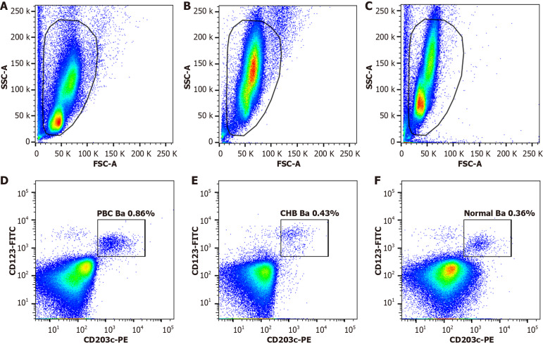
Methods: This single-center retrospective case-control study conducted at Guangdong Medical University Affiliated Hospital (China) between September 2019 and August 2024 enrolled 65 consecutive treatment-naïve patients with PBC (PBC group), 65 age- and sex-matched patients with chronic hepatitis B (CHB group), and 65 healthy controls (Normal group). Fourteen participants per group (subgroup) were randomly selected for flow cytometry analysis of basophil proportion, activation markers (CD203c and CD62 L mean fluorescence intensity), IL-6-positive basophils (IL-6+ basophils as a percentage of total basophils), and IL-17-positive T lymphocytes (CD3+CD4+IL-17+ cells) proportion among T cells. Data were analyzed using Kruskal-Wallis and χ2 tests as appropriate.
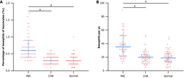
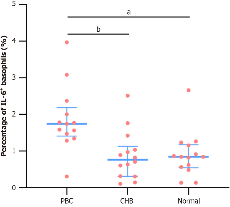
Results: Routine blood tests revealed significantly higher basophil counts and proportions in the PBC group compared to the CHB and Normal groups (P < 0.001 for both comparisons), with no significant differences between the CHB and Normal groups (P = 0.201). Flow cytometry revealed a higher basophil proportion in the PBC subgroup compared to the CHB (P = 0.011) and Normal subgroups (P < 0.001). The mean fluorescence intensity of CD203c on basophil surfaces was elevated in the PBC subgroup compared to the CHB (P = 0.032) and Normal subgroups (P = 0.039). The proportion of IL-6+ basophils was significantly higher in the PBC subgroup than in the CHB (P < 0.01) and Normal subgroups (P < 0.001). Similarly, the Th17 cell proportion was markedly elevated in the PBC compared to the CHB (P < 0.001) and Normal subgroups (P < 0.001).
Conclusion: Patients with PBC have increased peripheral basophil counts with enhanced activation. Activated basophils have increased IL-6 expression, which may indirectly induce Th17 cell proliferation and contribute to PBC pathogenesis.

 求助内容:
求助内容: 应助结果提醒方式:


