Hanan B Elajaili, Nathan Dee, Tanden Hovey, Autumn Canny, Georgina Amassah, Janelle Posey, George A Rinard, Joseph P Y Kao, Sandra S Eaton, Gareth R Eaton, Eva S Nozik
{"title":"电子顺磁共振检测小鼠急性肺损伤模型中的超氧化物。","authors":"Hanan B Elajaili, Nathan Dee, Tanden Hovey, Autumn Canny, Georgina Amassah, Janelle Posey, George A Rinard, Joseph P Y Kao, Sandra S Eaton, Gareth R Eaton, Eva S Nozik","doi":"10.1007/s44352-025-00014-1","DOIUrl":null,"url":null,"abstract":"<p><p>Superoxide (O<sub>2</sub> <sup>·-</sup>) production in an acute lung injury (ALI) murine model was detected by electron paramagnetic resonance (EPR) spectroscopy and imaging. Lung injury was induced in wild-type (WT) mice and transgenic (Tg) mice with lung-specific EC-SOD overexpression by lipopolysaccharide (LPS) administered intraperitoneally (IP) at a dose of 10 mg/kg. At 24 h after LPS treatment, mice were treated intraperitoneally and subcutaneously with the cyclic hydroxylamine probe, CMH, for superoxide measurements in the blood, or via intratracheal delivery (IT) with the cyclic hydroxylamine probes, CPH or DCP-AM-H, for lung cellular and mitochondrial superoxide detection. Blood was drawn one hour after CMH probe administration, while lungs were harvested five minutes following the administration of CPH or DCP-AM-H. Superoxide measurements in the blood by EPR were performed at X-band (~ 9.5 GHz). EPR images of isolated lungs were obtained by rapid-scan EPR at L-band (1 GHz). Inflammatory cell count, protein, and cell count in bronchoalveolar lavage fluid (BALF) were used to evaluate systemic inflammation and lung injury, respectively. Increased circulating neutrophils and monocytes indicate LPS-induced systemic inflammation. LPS-induced ALI was evidenced by increased alveolar protein and inflammatory cell count. In WT mice LPS increased superoxide in blood and increased lung cellular and mitochondrial superoxide, measured by EPR. In Tg mice with increased lung EC-SOD, blood superoxide increased; however, lung cellular and mitochondrial superoxide did not increase with LPS. These results show that EPR spectroscopy and imaging of excised lungs can detect superoxide production in a model of ALI and differentiate between cellular and mitochondrial superoxide. This provides essential new information as we showed that changes in lung superoxide does not always correlate with changes in blood superoxide levels. This is a significant step toward the ultimate goal of establishing a protocol for real-time monitoring of lung redox status in vivo, enabling disease risk stratification and guiding clinical research.</p><p><strong>Supplementary information: </strong>The online version contains supplementary material available at 10.1007/s44352-025-00014-1.</p>","PeriodicalId":520461,"journal":{"name":"Discover imaging","volume":"2 1","pages":"11"},"PeriodicalIF":0.0000,"publicationDate":"2025-01-01","publicationTypes":"Journal Article","fieldsOfStudy":null,"isOpenAccess":false,"openAccessPdf":"https://www.ncbi.nlm.nih.gov/pmc/articles/PMC12394321/pdf/","citationCount":"0","resultStr":"{\"title\":\"Electron paramagnetic resonance detection of superoxide in a murine model of acute lung injury.\",\"authors\":\"Hanan B Elajaili, Nathan Dee, Tanden Hovey, Autumn Canny, Georgina Amassah, Janelle Posey, George A Rinard, Joseph P Y Kao, Sandra S Eaton, Gareth R Eaton, Eva S Nozik\",\"doi\":\"10.1007/s44352-025-00014-1\",\"DOIUrl\":null,\"url\":null,\"abstract\":\"<p><p>Superoxide (O<sub>2</sub> <sup>·-</sup>) production in an acute lung injury (ALI) murine model was detected by electron paramagnetic resonance (EPR) spectroscopy and imaging. Lung injury was induced in wild-type (WT) mice and transgenic (Tg) mice with lung-specific EC-SOD overexpression by lipopolysaccharide (LPS) administered intraperitoneally (IP) at a dose of 10 mg/kg. At 24 h after LPS treatment, mice were treated intraperitoneally and subcutaneously with the cyclic hydroxylamine probe, CMH, for superoxide measurements in the blood, or via intratracheal delivery (IT) with the cyclic hydroxylamine probes, CPH or DCP-AM-H, for lung cellular and mitochondrial superoxide detection. Blood was drawn one hour after CMH probe administration, while lungs were harvested five minutes following the administration of CPH or DCP-AM-H. Superoxide measurements in the blood by EPR were performed at X-band (~ 9.5 GHz). EPR images of isolated lungs were obtained by rapid-scan EPR at L-band (1 GHz). Inflammatory cell count, protein, and cell count in bronchoalveolar lavage fluid (BALF) were used to evaluate systemic inflammation and lung injury, respectively. Increased circulating neutrophils and monocytes indicate LPS-induced systemic inflammation. LPS-induced ALI was evidenced by increased alveolar protein and inflammatory cell count. In WT mice LPS increased superoxide in blood and increased lung cellular and mitochondrial superoxide, measured by EPR. In Tg mice with increased lung EC-SOD, blood superoxide increased; however, lung cellular and mitochondrial superoxide did not increase with LPS. These results show that EPR spectroscopy and imaging of excised lungs can detect superoxide production in a model of ALI and differentiate between cellular and mitochondrial superoxide. This provides essential new information as we showed that changes in lung superoxide does not always correlate with changes in blood superoxide levels. This is a significant step toward the ultimate goal of establishing a protocol for real-time monitoring of lung redox status in vivo, enabling disease risk stratification and guiding clinical research.</p><p><strong>Supplementary information: </strong>The online version contains supplementary material available at 10.1007/s44352-025-00014-1.</p>\",\"PeriodicalId\":520461,\"journal\":{\"name\":\"Discover imaging\",\"volume\":\"2 1\",\"pages\":\"11\"},\"PeriodicalIF\":0.0000,\"publicationDate\":\"2025-01-01\",\"publicationTypes\":\"Journal Article\",\"fieldsOfStudy\":null,\"isOpenAccess\":false,\"openAccessPdf\":\"https://www.ncbi.nlm.nih.gov/pmc/articles/PMC12394321/pdf/\",\"citationCount\":\"0\",\"resultStr\":null,\"platform\":\"Semanticscholar\",\"paperid\":null,\"PeriodicalName\":\"Discover imaging\",\"FirstCategoryId\":\"1085\",\"ListUrlMain\":\"https://doi.org/10.1007/s44352-025-00014-1\",\"RegionNum\":0,\"RegionCategory\":null,\"ArticlePicture\":[],\"TitleCN\":null,\"AbstractTextCN\":null,\"PMCID\":null,\"EPubDate\":\"2025/8/28 0:00:00\",\"PubModel\":\"Epub\",\"JCR\":\"\",\"JCRName\":\"\",\"Score\":null,\"Total\":0}","platform":"Semanticscholar","paperid":null,"PeriodicalName":"Discover imaging","FirstCategoryId":"1085","ListUrlMain":"https://doi.org/10.1007/s44352-025-00014-1","RegionNum":0,"RegionCategory":null,"ArticlePicture":[],"TitleCN":null,"AbstractTextCN":null,"PMCID":null,"EPubDate":"2025/8/28 0:00:00","PubModel":"Epub","JCR":"","JCRName":"","Score":null,"Total":0}
Electron paramagnetic resonance detection of superoxide in a murine model of acute lung injury.
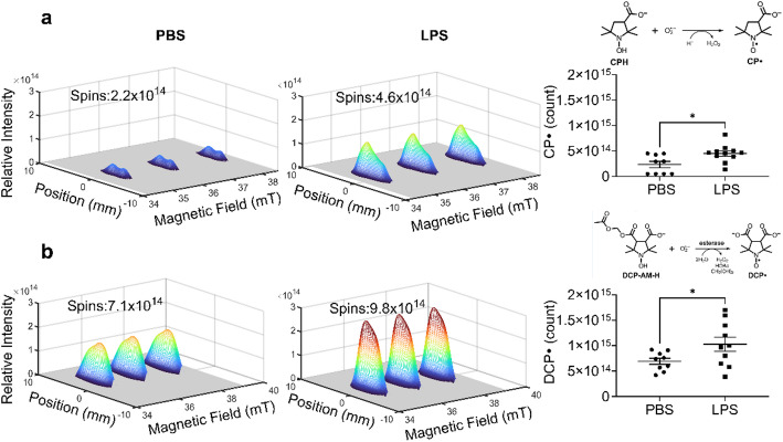
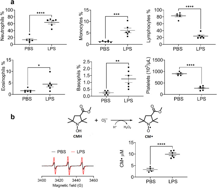
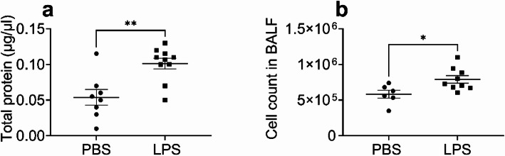
Superoxide (O2·-) production in an acute lung injury (ALI) murine model was detected by electron paramagnetic resonance (EPR) spectroscopy and imaging. Lung injury was induced in wild-type (WT) mice and transgenic (Tg) mice with lung-specific EC-SOD overexpression by lipopolysaccharide (LPS) administered intraperitoneally (IP) at a dose of 10 mg/kg. At 24 h after LPS treatment, mice were treated intraperitoneally and subcutaneously with the cyclic hydroxylamine probe, CMH, for superoxide measurements in the blood, or via intratracheal delivery (IT) with the cyclic hydroxylamine probes, CPH or DCP-AM-H, for lung cellular and mitochondrial superoxide detection. Blood was drawn one hour after CMH probe administration, while lungs were harvested five minutes following the administration of CPH or DCP-AM-H. Superoxide measurements in the blood by EPR were performed at X-band (~ 9.5 GHz). EPR images of isolated lungs were obtained by rapid-scan EPR at L-band (1 GHz). Inflammatory cell count, protein, and cell count in bronchoalveolar lavage fluid (BALF) were used to evaluate systemic inflammation and lung injury, respectively. Increased circulating neutrophils and monocytes indicate LPS-induced systemic inflammation. LPS-induced ALI was evidenced by increased alveolar protein and inflammatory cell count. In WT mice LPS increased superoxide in blood and increased lung cellular and mitochondrial superoxide, measured by EPR. In Tg mice with increased lung EC-SOD, blood superoxide increased; however, lung cellular and mitochondrial superoxide did not increase with LPS. These results show that EPR spectroscopy and imaging of excised lungs can detect superoxide production in a model of ALI and differentiate between cellular and mitochondrial superoxide. This provides essential new information as we showed that changes in lung superoxide does not always correlate with changes in blood superoxide levels. This is a significant step toward the ultimate goal of establishing a protocol for real-time monitoring of lung redox status in vivo, enabling disease risk stratification and guiding clinical research.
Supplementary information: The online version contains supplementary material available at 10.1007/s44352-025-00014-1.

 求助内容:
求助内容: 应助结果提醒方式:


