{"title":"保留颞浅动脉的翼点头皮皮瓣颞浅动脉主干及顶支的走行:尸体及临床研究。","authors":"Nattamon Suanchan, Kitiporn Sriamornrattanakul, Thirawass Phumyoo","doi":"10.1055/s-0045-1809324","DOIUrl":null,"url":null,"abstract":"<p><strong>Background: </strong>The pterional incision is usually performed near the course of the superficial temporal artery (STA), which carries a risk of injury to a branch or even the main trunk of the STA (mSTA). In this study, we assessed the usual course of the mSTA and its parietal branch of the STA (pSTA) and evaluated the efficacy of a modified pterional scalp incision for the preservation of all STA branches.</p><p><strong>Materials and methods: </strong>Sixteen sides of cadaveric heads were dissected to study the location and paths of the mSTA and pSTA in the vicinity of the ear cartilage and the oculomeatal (OM) line. We also performed a clinical study of 31 patients who underwent pterional craniotomy using the modified pterional scalp incision. Postoperative STA preservation was retrospectively evaluated.</p><p><strong>Results: </strong>The mean distances between the mSTA and the anterior edge of the ear cartilage were 0.5 and 0.6 mm. The mean angle of the pSTA axis was 88.8 degrees (range 75-95 degrees) from the OM line. Among the patients treated using the modified pterional scalp incision, the mSTA, the pSTA, and the frontal branch of the STA (fSTA) were preserved within the scalp flap in 93.5, 96.7, and 88.9%, respectively.</p><p><strong>Conclusion: </strong>The mSTA was found to pass very close to the ear cartilage, while the axis of pSTA coursed approximately perpendicular to the OM line. To preserve all branches of the STA, the pterional skin incision should begin just anterior to the ear cartilage and then curve slightly to the posterior above the pinna.</p>","PeriodicalId":94300,"journal":{"name":"Asian journal of neurosurgery","volume":"20 3","pages":"581-589"},"PeriodicalIF":0.0000,"publicationDate":"2025-05-20","publicationTypes":"Journal Article","fieldsOfStudy":null,"isOpenAccess":false,"openAccessPdf":"https://www.ncbi.nlm.nih.gov/pmc/articles/PMC12370334/pdf/","citationCount":"0","resultStr":"{\"title\":\"The Course of the Main Trunk and Parietal Branch of the Superficial Temporal Artery for a Pterional Scalp Flap with Superficial Temporal Artery Preservation: Cadaveric and Clinical Study.\",\"authors\":\"Nattamon Suanchan, Kitiporn Sriamornrattanakul, Thirawass Phumyoo\",\"doi\":\"10.1055/s-0045-1809324\",\"DOIUrl\":null,\"url\":null,\"abstract\":\"<p><strong>Background: </strong>The pterional incision is usually performed near the course of the superficial temporal artery (STA), which carries a risk of injury to a branch or even the main trunk of the STA (mSTA). In this study, we assessed the usual course of the mSTA and its parietal branch of the STA (pSTA) and evaluated the efficacy of a modified pterional scalp incision for the preservation of all STA branches.</p><p><strong>Materials and methods: </strong>Sixteen sides of cadaveric heads were dissected to study the location and paths of the mSTA and pSTA in the vicinity of the ear cartilage and the oculomeatal (OM) line. We also performed a clinical study of 31 patients who underwent pterional craniotomy using the modified pterional scalp incision. Postoperative STA preservation was retrospectively evaluated.</p><p><strong>Results: </strong>The mean distances between the mSTA and the anterior edge of the ear cartilage were 0.5 and 0.6 mm. The mean angle of the pSTA axis was 88.8 degrees (range 75-95 degrees) from the OM line. Among the patients treated using the modified pterional scalp incision, the mSTA, the pSTA, and the frontal branch of the STA (fSTA) were preserved within the scalp flap in 93.5, 96.7, and 88.9%, respectively.</p><p><strong>Conclusion: </strong>The mSTA was found to pass very close to the ear cartilage, while the axis of pSTA coursed approximately perpendicular to the OM line. To preserve all branches of the STA, the pterional skin incision should begin just anterior to the ear cartilage and then curve slightly to the posterior above the pinna.</p>\",\"PeriodicalId\":94300,\"journal\":{\"name\":\"Asian journal of neurosurgery\",\"volume\":\"20 3\",\"pages\":\"581-589\"},\"PeriodicalIF\":0.0000,\"publicationDate\":\"2025-05-20\",\"publicationTypes\":\"Journal Article\",\"fieldsOfStudy\":null,\"isOpenAccess\":false,\"openAccessPdf\":\"https://www.ncbi.nlm.nih.gov/pmc/articles/PMC12370334/pdf/\",\"citationCount\":\"0\",\"resultStr\":null,\"platform\":\"Semanticscholar\",\"paperid\":null,\"PeriodicalName\":\"Asian journal of neurosurgery\",\"FirstCategoryId\":\"1085\",\"ListUrlMain\":\"https://doi.org/10.1055/s-0045-1809324\",\"RegionNum\":0,\"RegionCategory\":null,\"ArticlePicture\":[],\"TitleCN\":null,\"AbstractTextCN\":null,\"PMCID\":null,\"EPubDate\":\"2025/9/1 0:00:00\",\"PubModel\":\"eCollection\",\"JCR\":\"\",\"JCRName\":\"\",\"Score\":null,\"Total\":0}","platform":"Semanticscholar","paperid":null,"PeriodicalName":"Asian journal of neurosurgery","FirstCategoryId":"1085","ListUrlMain":"https://doi.org/10.1055/s-0045-1809324","RegionNum":0,"RegionCategory":null,"ArticlePicture":[],"TitleCN":null,"AbstractTextCN":null,"PMCID":null,"EPubDate":"2025/9/1 0:00:00","PubModel":"eCollection","JCR":"","JCRName":"","Score":null,"Total":0}
The Course of the Main Trunk and Parietal Branch of the Superficial Temporal Artery for a Pterional Scalp Flap with Superficial Temporal Artery Preservation: Cadaveric and Clinical Study.
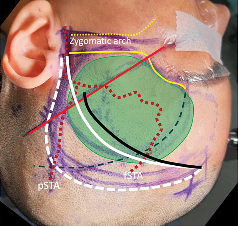
Background: The pterional incision is usually performed near the course of the superficial temporal artery (STA), which carries a risk of injury to a branch or even the main trunk of the STA (mSTA). In this study, we assessed the usual course of the mSTA and its parietal branch of the STA (pSTA) and evaluated the efficacy of a modified pterional scalp incision for the preservation of all STA branches.
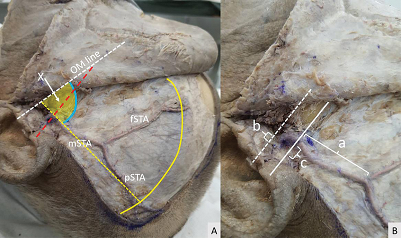
Materials and methods: Sixteen sides of cadaveric heads were dissected to study the location and paths of the mSTA and pSTA in the vicinity of the ear cartilage and the oculomeatal (OM) line. We also performed a clinical study of 31 patients who underwent pterional craniotomy using the modified pterional scalp incision. Postoperative STA preservation was retrospectively evaluated.
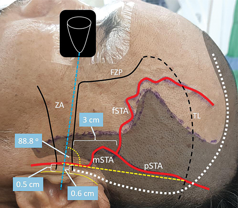
Results: The mean distances between the mSTA and the anterior edge of the ear cartilage were 0.5 and 0.6 mm. The mean angle of the pSTA axis was 88.8 degrees (range 75-95 degrees) from the OM line. Among the patients treated using the modified pterional scalp incision, the mSTA, the pSTA, and the frontal branch of the STA (fSTA) were preserved within the scalp flap in 93.5, 96.7, and 88.9%, respectively.
Conclusion: The mSTA was found to pass very close to the ear cartilage, while the axis of pSTA coursed approximately perpendicular to the OM line. To preserve all branches of the STA, the pterional skin incision should begin just anterior to the ear cartilage and then curve slightly to the posterior above the pinna.

 求助内容:
求助内容: 应助结果提醒方式:


