Mark D Johnson, Pradyumna Elavarthi, Seth Street, Samer S Hoz, Anca L Ralescu, Charles J Prestigiacomo
{"title":"使用临床和影像学变量预测颅内动脉瘤破裂状态与机器学习。","authors":"Mark D Johnson, Pradyumna Elavarthi, Seth Street, Samer S Hoz, Anca L Ralescu, Charles J Prestigiacomo","doi":"10.25259/SNI_498_2025","DOIUrl":null,"url":null,"abstract":"<p><strong>Background: </strong>With excitement in the medical community around artificial intelligence, machine learning (ML) techniques have been applied to correlate clinical and radiographic variables with intracranial aneurysm (IA) rupture status. In this study, we applied various ML techniques, including random forest (RF), XGBoost (XGB), support vector machines (SVM), and multi-layer perceptron (MLP), to predict IA rupture status.</p><p><strong>Methods: </strong>The dataset consisted of 178 IAs each with 53 clinical and radiographic features for analysis. We removed features with high correlation (>0.8) with respect to the target variable to reduce redundancy. We applied grid search to fine-tune the hyperparameters for each model. Each model was evaluated across five iterations of 5-fold cross-validation. Overall performance metrics (accuracy, precision, recall, and F1-score) were extracted. The Wilcoxon signed-rank test was used to compare the area under the curve (AUC) scores between models.</p><p><strong>Results: </strong>The most common locations were internal carotid artery (42), anterior communicating artery (41), middle cerebral artery (32), and posterior communicating artery (25). The AUC for the RF (0.85) and XGB (0.76) models were significantly higher than those for the SVM (0.69) and MLP (0.65) models (<i>P</i> < 0.05). There was no statistical difference in accuracy between RF and XBG models (<i>P</i> = 0.144). Fractal dimension ranked as the most important feature for model performance across all models. Three-dimensional (3D) shape features made up 8 of the 15 most important features driving model performance.</p><p><strong>Conclusion: </strong>Among the models, RF achieved the highest accuracy (85%) with balanced precision and recall. Across models 3D geometric features drove model performance, highlighting the importance of these features in predicting rupture status.</p>","PeriodicalId":94217,"journal":{"name":"Surgical neurology international","volume":"16 ","pages":"298"},"PeriodicalIF":0.0000,"publicationDate":"2025-07-18","publicationTypes":"Journal Article","fieldsOfStudy":null,"isOpenAccess":false,"openAccessPdf":"https://www.ncbi.nlm.nih.gov/pmc/articles/PMC12361647/pdf/","citationCount":"0","resultStr":"{\"title\":\"Using clinical and radiographic variables to predict intracranial aneurysm rupture status with machine learning.\",\"authors\":\"Mark D Johnson, Pradyumna Elavarthi, Seth Street, Samer S Hoz, Anca L Ralescu, Charles J Prestigiacomo\",\"doi\":\"10.25259/SNI_498_2025\",\"DOIUrl\":null,\"url\":null,\"abstract\":\"<p><strong>Background: </strong>With excitement in the medical community around artificial intelligence, machine learning (ML) techniques have been applied to correlate clinical and radiographic variables with intracranial aneurysm (IA) rupture status. In this study, we applied various ML techniques, including random forest (RF), XGBoost (XGB), support vector machines (SVM), and multi-layer perceptron (MLP), to predict IA rupture status.</p><p><strong>Methods: </strong>The dataset consisted of 178 IAs each with 53 clinical and radiographic features for analysis. We removed features with high correlation (>0.8) with respect to the target variable to reduce redundancy. We applied grid search to fine-tune the hyperparameters for each model. Each model was evaluated across five iterations of 5-fold cross-validation. Overall performance metrics (accuracy, precision, recall, and F1-score) were extracted. The Wilcoxon signed-rank test was used to compare the area under the curve (AUC) scores between models.</p><p><strong>Results: </strong>The most common locations were internal carotid artery (42), anterior communicating artery (41), middle cerebral artery (32), and posterior communicating artery (25). The AUC for the RF (0.85) and XGB (0.76) models were significantly higher than those for the SVM (0.69) and MLP (0.65) models (<i>P</i> < 0.05). There was no statistical difference in accuracy between RF and XBG models (<i>P</i> = 0.144). Fractal dimension ranked as the most important feature for model performance across all models. Three-dimensional (3D) shape features made up 8 of the 15 most important features driving model performance.</p><p><strong>Conclusion: </strong>Among the models, RF achieved the highest accuracy (85%) with balanced precision and recall. Across models 3D geometric features drove model performance, highlighting the importance of these features in predicting rupture status.</p>\",\"PeriodicalId\":94217,\"journal\":{\"name\":\"Surgical neurology international\",\"volume\":\"16 \",\"pages\":\"298\"},\"PeriodicalIF\":0.0000,\"publicationDate\":\"2025-07-18\",\"publicationTypes\":\"Journal Article\",\"fieldsOfStudy\":null,\"isOpenAccess\":false,\"openAccessPdf\":\"https://www.ncbi.nlm.nih.gov/pmc/articles/PMC12361647/pdf/\",\"citationCount\":\"0\",\"resultStr\":null,\"platform\":\"Semanticscholar\",\"paperid\":null,\"PeriodicalName\":\"Surgical neurology international\",\"FirstCategoryId\":\"1085\",\"ListUrlMain\":\"https://doi.org/10.25259/SNI_498_2025\",\"RegionNum\":0,\"RegionCategory\":null,\"ArticlePicture\":[],\"TitleCN\":null,\"AbstractTextCN\":null,\"PMCID\":null,\"EPubDate\":\"2025/1/1 0:00:00\",\"PubModel\":\"eCollection\",\"JCR\":\"\",\"JCRName\":\"\",\"Score\":null,\"Total\":0}","platform":"Semanticscholar","paperid":null,"PeriodicalName":"Surgical neurology international","FirstCategoryId":"1085","ListUrlMain":"https://doi.org/10.25259/SNI_498_2025","RegionNum":0,"RegionCategory":null,"ArticlePicture":[],"TitleCN":null,"AbstractTextCN":null,"PMCID":null,"EPubDate":"2025/1/1 0:00:00","PubModel":"eCollection","JCR":"","JCRName":"","Score":null,"Total":0}
Using clinical and radiographic variables to predict intracranial aneurysm rupture status with machine learning.
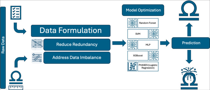
Background: With excitement in the medical community around artificial intelligence, machine learning (ML) techniques have been applied to correlate clinical and radiographic variables with intracranial aneurysm (IA) rupture status. In this study, we applied various ML techniques, including random forest (RF), XGBoost (XGB), support vector machines (SVM), and multi-layer perceptron (MLP), to predict IA rupture status.
Methods: The dataset consisted of 178 IAs each with 53 clinical and radiographic features for analysis. We removed features with high correlation (>0.8) with respect to the target variable to reduce redundancy. We applied grid search to fine-tune the hyperparameters for each model. Each model was evaluated across five iterations of 5-fold cross-validation. Overall performance metrics (accuracy, precision, recall, and F1-score) were extracted. The Wilcoxon signed-rank test was used to compare the area under the curve (AUC) scores between models.
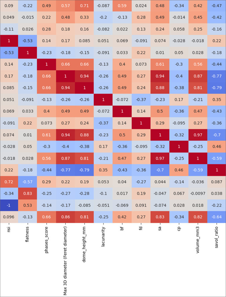
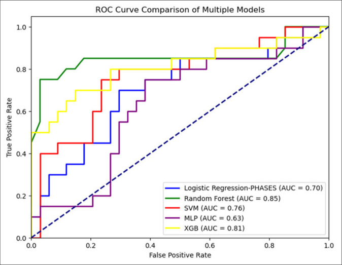
Results: The most common locations were internal carotid artery (42), anterior communicating artery (41), middle cerebral artery (32), and posterior communicating artery (25). The AUC for the RF (0.85) and XGB (0.76) models were significantly higher than those for the SVM (0.69) and MLP (0.65) models (P < 0.05). There was no statistical difference in accuracy between RF and XBG models (P = 0.144). Fractal dimension ranked as the most important feature for model performance across all models. Three-dimensional (3D) shape features made up 8 of the 15 most important features driving model performance.
Conclusion: Among the models, RF achieved the highest accuracy (85%) with balanced precision and recall. Across models 3D geometric features drove model performance, highlighting the importance of these features in predicting rupture status.

 求助内容:
求助内容: 应助结果提醒方式:


