{"title":"Meckel穴症状性假瘤模拟三叉神经鞘瘤。","authors":"Tomoya Sofue, Megumi Chatani, Yoshihiro Kuga, Hiroyuki Ohnishi","doi":"10.25259/SNI_447_2025","DOIUrl":null,"url":null,"abstract":"<p><strong>Background: </strong>Inflammatory pseudotumor is non-neoplastic lesions that form tumor-like masses, typically occurring in the lungs and orbits, with rare involvement of intracranial regions such as the Meckel cave. While neuroimaging can assist in differential diagnosis, definitive diagnosis may remain challenging. Some cases are associated with immunoglobulin G4 (IgG4)-related disease, highlighting the importance of histopathological confirmation to guide appropriate management.</p><p><strong>Case description: </strong>A case of a 42-year-old woman presented with facial numbness and headache. Magnetic resonance imaging revealed a 25-mm enhancing mass in the left Meckel cave, initially suspected to be a trigeminal schwannoma. Craniotomy and tumor resection were performed. Intraoperative findings and rapid pathology indicated marked inflammatory cell infiltration without features of schwannoma. The final diagnosis was inflammatory pseudotumor, with no evidence of IgG4-related disease or malignancy. Postoperative symptoms improved, and only a short course of steroids was administered.</p><p><strong>Conclusion: </strong>This case underscores the diagnostic difficulty of distinguishing inflammatory pseudotumor from neoplastic lesions in skull base regions. Surgical biopsy or resection is critical for definitive diagnosis, especially in cases with progressive symptoms. Individualized treatment planning based on disease activity and neurological impact remains essential due to the absence of standardized treatment guidelines.</p>","PeriodicalId":94217,"journal":{"name":"Surgical neurology international","volume":"16 ","pages":"280"},"PeriodicalIF":0.0000,"publicationDate":"2025-07-11","publicationTypes":"Journal Article","fieldsOfStudy":null,"isOpenAccess":false,"openAccessPdf":"https://www.ncbi.nlm.nih.gov/pmc/articles/PMC12361671/pdf/","citationCount":"0","resultStr":"{\"title\":\"Symptomatic pseudotumor in Meckel cave mimics trigeminal schwannoma.\",\"authors\":\"Tomoya Sofue, Megumi Chatani, Yoshihiro Kuga, Hiroyuki Ohnishi\",\"doi\":\"10.25259/SNI_447_2025\",\"DOIUrl\":null,\"url\":null,\"abstract\":\"<p><strong>Background: </strong>Inflammatory pseudotumor is non-neoplastic lesions that form tumor-like masses, typically occurring in the lungs and orbits, with rare involvement of intracranial regions such as the Meckel cave. While neuroimaging can assist in differential diagnosis, definitive diagnosis may remain challenging. Some cases are associated with immunoglobulin G4 (IgG4)-related disease, highlighting the importance of histopathological confirmation to guide appropriate management.</p><p><strong>Case description: </strong>A case of a 42-year-old woman presented with facial numbness and headache. Magnetic resonance imaging revealed a 25-mm enhancing mass in the left Meckel cave, initially suspected to be a trigeminal schwannoma. Craniotomy and tumor resection were performed. Intraoperative findings and rapid pathology indicated marked inflammatory cell infiltration without features of schwannoma. The final diagnosis was inflammatory pseudotumor, with no evidence of IgG4-related disease or malignancy. Postoperative symptoms improved, and only a short course of steroids was administered.</p><p><strong>Conclusion: </strong>This case underscores the diagnostic difficulty of distinguishing inflammatory pseudotumor from neoplastic lesions in skull base regions. Surgical biopsy or resection is critical for definitive diagnosis, especially in cases with progressive symptoms. Individualized treatment planning based on disease activity and neurological impact remains essential due to the absence of standardized treatment guidelines.</p>\",\"PeriodicalId\":94217,\"journal\":{\"name\":\"Surgical neurology international\",\"volume\":\"16 \",\"pages\":\"280\"},\"PeriodicalIF\":0.0000,\"publicationDate\":\"2025-07-11\",\"publicationTypes\":\"Journal Article\",\"fieldsOfStudy\":null,\"isOpenAccess\":false,\"openAccessPdf\":\"https://www.ncbi.nlm.nih.gov/pmc/articles/PMC12361671/pdf/\",\"citationCount\":\"0\",\"resultStr\":null,\"platform\":\"Semanticscholar\",\"paperid\":null,\"PeriodicalName\":\"Surgical neurology international\",\"FirstCategoryId\":\"1085\",\"ListUrlMain\":\"https://doi.org/10.25259/SNI_447_2025\",\"RegionNum\":0,\"RegionCategory\":null,\"ArticlePicture\":[],\"TitleCN\":null,\"AbstractTextCN\":null,\"PMCID\":null,\"EPubDate\":\"2025/1/1 0:00:00\",\"PubModel\":\"eCollection\",\"JCR\":\"\",\"JCRName\":\"\",\"Score\":null,\"Total\":0}","platform":"Semanticscholar","paperid":null,"PeriodicalName":"Surgical neurology international","FirstCategoryId":"1085","ListUrlMain":"https://doi.org/10.25259/SNI_447_2025","RegionNum":0,"RegionCategory":null,"ArticlePicture":[],"TitleCN":null,"AbstractTextCN":null,"PMCID":null,"EPubDate":"2025/1/1 0:00:00","PubModel":"eCollection","JCR":"","JCRName":"","Score":null,"Total":0}
Symptomatic pseudotumor in Meckel cave mimics trigeminal schwannoma.
Background: Inflammatory pseudotumor is non-neoplastic lesions that form tumor-like masses, typically occurring in the lungs and orbits, with rare involvement of intracranial regions such as the Meckel cave. While neuroimaging can assist in differential diagnosis, definitive diagnosis may remain challenging. Some cases are associated with immunoglobulin G4 (IgG4)-related disease, highlighting the importance of histopathological confirmation to guide appropriate management.
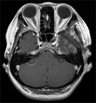
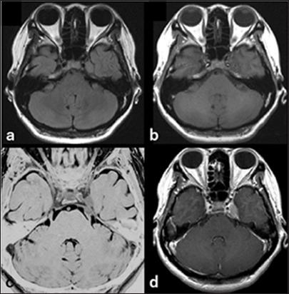
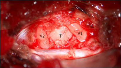
Case description: A case of a 42-year-old woman presented with facial numbness and headache. Magnetic resonance imaging revealed a 25-mm enhancing mass in the left Meckel cave, initially suspected to be a trigeminal schwannoma. Craniotomy and tumor resection were performed. Intraoperative findings and rapid pathology indicated marked inflammatory cell infiltration without features of schwannoma. The final diagnosis was inflammatory pseudotumor, with no evidence of IgG4-related disease or malignancy. Postoperative symptoms improved, and only a short course of steroids was administered.
Conclusion: This case underscores the diagnostic difficulty of distinguishing inflammatory pseudotumor from neoplastic lesions in skull base regions. Surgical biopsy or resection is critical for definitive diagnosis, especially in cases with progressive symptoms. Individualized treatment planning based on disease activity and neurological impact remains essential due to the absence of standardized treatment guidelines.

 求助内容:
求助内容: 应助结果提醒方式:


