{"title":"海绵血管瘤硬膜外T3-T5出血致截瘫。","authors":"Ghassen Gader, Emna Mzoughi, Olfa Faten, Alia Zehani, Mouna Rkhami, Ihsèn Zammel","doi":"10.25259/SNI_504_2025","DOIUrl":null,"url":null,"abstract":"<p><strong>Background: </strong>Epidural cavernous hemangiomas (ECHs) are rare, benign vascular malformations. They account for 4% of all epidural lesions, and 5-12% of all spinal vascular malformations. Acute hemorrhages into extra-hepatic components may cause the acute onset of major neurological deficits.</p><p><strong>Case description: </strong>A 70-year-old male presented with 3 months of progressive paraparesis. The magnetic resonance showed a compressive epidural T3-T5 lesion that was successfully removed. The lesion proved histopathologically to be a benign cavernous angioma.</p><p><strong>Conclusion: </strong>For patients presenting with progressive myelopathy, early diagnosis and timely surgical resection of benign spinal epidural cavernous angiomas are critical to optimize outcomes.</p>","PeriodicalId":94217,"journal":{"name":"Surgical neurology international","volume":"16 ","pages":"303"},"PeriodicalIF":0.0000,"publicationDate":"2025-07-25","publicationTypes":"Journal Article","fieldsOfStudy":null,"isOpenAccess":false,"openAccessPdf":"https://www.ncbi.nlm.nih.gov/pmc/articles/PMC12361715/pdf/","citationCount":"0","resultStr":"{\"title\":\"Paraplegia due to epidural T3-T5 hemorrhage of cavernous hemangioma.\",\"authors\":\"Ghassen Gader, Emna Mzoughi, Olfa Faten, Alia Zehani, Mouna Rkhami, Ihsèn Zammel\",\"doi\":\"10.25259/SNI_504_2025\",\"DOIUrl\":null,\"url\":null,\"abstract\":\"<p><strong>Background: </strong>Epidural cavernous hemangiomas (ECHs) are rare, benign vascular malformations. They account for 4% of all epidural lesions, and 5-12% of all spinal vascular malformations. Acute hemorrhages into extra-hepatic components may cause the acute onset of major neurological deficits.</p><p><strong>Case description: </strong>A 70-year-old male presented with 3 months of progressive paraparesis. The magnetic resonance showed a compressive epidural T3-T5 lesion that was successfully removed. The lesion proved histopathologically to be a benign cavernous angioma.</p><p><strong>Conclusion: </strong>For patients presenting with progressive myelopathy, early diagnosis and timely surgical resection of benign spinal epidural cavernous angiomas are critical to optimize outcomes.</p>\",\"PeriodicalId\":94217,\"journal\":{\"name\":\"Surgical neurology international\",\"volume\":\"16 \",\"pages\":\"303\"},\"PeriodicalIF\":0.0000,\"publicationDate\":\"2025-07-25\",\"publicationTypes\":\"Journal Article\",\"fieldsOfStudy\":null,\"isOpenAccess\":false,\"openAccessPdf\":\"https://www.ncbi.nlm.nih.gov/pmc/articles/PMC12361715/pdf/\",\"citationCount\":\"0\",\"resultStr\":null,\"platform\":\"Semanticscholar\",\"paperid\":null,\"PeriodicalName\":\"Surgical neurology international\",\"FirstCategoryId\":\"1085\",\"ListUrlMain\":\"https://doi.org/10.25259/SNI_504_2025\",\"RegionNum\":0,\"RegionCategory\":null,\"ArticlePicture\":[],\"TitleCN\":null,\"AbstractTextCN\":null,\"PMCID\":null,\"EPubDate\":\"2025/1/1 0:00:00\",\"PubModel\":\"eCollection\",\"JCR\":\"\",\"JCRName\":\"\",\"Score\":null,\"Total\":0}","platform":"Semanticscholar","paperid":null,"PeriodicalName":"Surgical neurology international","FirstCategoryId":"1085","ListUrlMain":"https://doi.org/10.25259/SNI_504_2025","RegionNum":0,"RegionCategory":null,"ArticlePicture":[],"TitleCN":null,"AbstractTextCN":null,"PMCID":null,"EPubDate":"2025/1/1 0:00:00","PubModel":"eCollection","JCR":"","JCRName":"","Score":null,"Total":0}
Paraplegia due to epidural T3-T5 hemorrhage of cavernous hemangioma.
Background: Epidural cavernous hemangiomas (ECHs) are rare, benign vascular malformations. They account for 4% of all epidural lesions, and 5-12% of all spinal vascular malformations. Acute hemorrhages into extra-hepatic components may cause the acute onset of major neurological deficits.
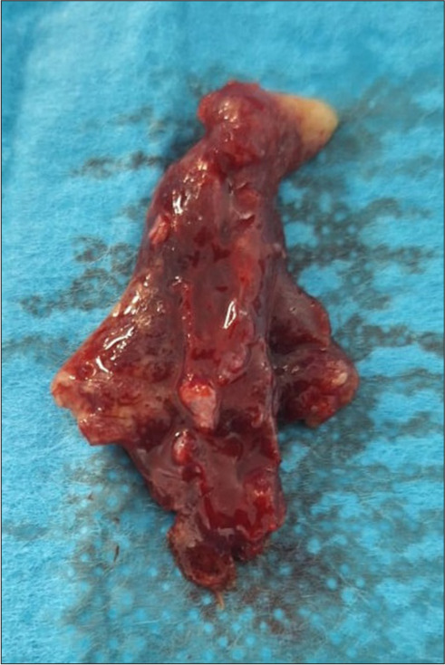
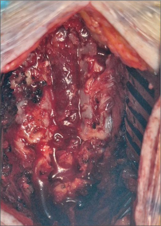
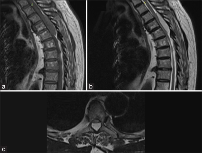
Case description: A 70-year-old male presented with 3 months of progressive paraparesis. The magnetic resonance showed a compressive epidural T3-T5 lesion that was successfully removed. The lesion proved histopathologically to be a benign cavernous angioma.
Conclusion: For patients presenting with progressive myelopathy, early diagnosis and timely surgical resection of benign spinal epidural cavernous angiomas are critical to optimize outcomes.

 求助内容:
求助内容: 应助结果提醒方式:


