Ahoud Alharbi, Ali Alassiri, Ali Alkhaibary, Saad AlQahatani
{"title":"青少年多形性低级别神经上皮肿瘤。","authors":"Ahoud Alharbi, Ali Alassiri, Ali Alkhaibary, Saad AlQahatani","doi":"10.25259/SNI_1061_2024","DOIUrl":null,"url":null,"abstract":"<p><strong>Background: </strong>Polymorphous low-grade neuroepithelial tumor of the young (PLNTY) is a newly recognized entity first described in 2017. This article reports the clinical, radiological, histological, and molecular characteristics of PLNTY diagnosed in a young female.</p><p><strong>Case description: </strong>A 20-year-old female, not known to have any medical illness, presented to the emergency department with speech arrest that progressed to generalized tonic-clonic seizures. Brain magnetic resonance imaging showed a T2 hyperintense nonenhancing cortical-based heterogeneous mass in the left medial temporal lobe and adjacent inferior temporal gyrus measuring 2.7 × 2 × 2.5 cm with no susceptibility signal or diffusion restriction. Electroencephalogram (EEG) showed left temporal epileptiform discharges. She underwent left frontotemporal craniotomy and tumor resection. The light microscopic examination of the tumor revealed a low-grade infiltrative neoplasm. There was a classical appearance of round cells with perinuclear halo, immunopositive for olig-2, glial fibrillary acidic protein (<i>GFAP)</i>, and cluster of differentiation-34. There was B-Raf protooncogene, serine/threonine kinase (<i>BRA</i>F) c.1799T>A (p.V600E) point mutation and absence of isocitrate dehydrogenase (<i>IDH)</i> 1 and 2 hotspot mutations. The overall findings were diagnostic of PLNTY.</p><p><strong>Conclusion: </strong>This article reports an additional case of PLNTY, a newly defined central nervous system tumor entity, describing its clinical, radiological, histological, and molecular features.</p>","PeriodicalId":94217,"journal":{"name":"Surgical neurology international","volume":"16 ","pages":"300"},"PeriodicalIF":0.0000,"publicationDate":"2025-07-25","publicationTypes":"Journal Article","fieldsOfStudy":null,"isOpenAccess":false,"openAccessPdf":"https://www.ncbi.nlm.nih.gov/pmc/articles/PMC12361669/pdf/","citationCount":"0","resultStr":"{\"title\":\"Polymorphous low-grade neuroepithelial tumor of the young.\",\"authors\":\"Ahoud Alharbi, Ali Alassiri, Ali Alkhaibary, Saad AlQahatani\",\"doi\":\"10.25259/SNI_1061_2024\",\"DOIUrl\":null,\"url\":null,\"abstract\":\"<p><strong>Background: </strong>Polymorphous low-grade neuroepithelial tumor of the young (PLNTY) is a newly recognized entity first described in 2017. This article reports the clinical, radiological, histological, and molecular characteristics of PLNTY diagnosed in a young female.</p><p><strong>Case description: </strong>A 20-year-old female, not known to have any medical illness, presented to the emergency department with speech arrest that progressed to generalized tonic-clonic seizures. Brain magnetic resonance imaging showed a T2 hyperintense nonenhancing cortical-based heterogeneous mass in the left medial temporal lobe and adjacent inferior temporal gyrus measuring 2.7 × 2 × 2.5 cm with no susceptibility signal or diffusion restriction. Electroencephalogram (EEG) showed left temporal epileptiform discharges. She underwent left frontotemporal craniotomy and tumor resection. The light microscopic examination of the tumor revealed a low-grade infiltrative neoplasm. There was a classical appearance of round cells with perinuclear halo, immunopositive for olig-2, glial fibrillary acidic protein (<i>GFAP)</i>, and cluster of differentiation-34. There was B-Raf protooncogene, serine/threonine kinase (<i>BRA</i>F) c.1799T>A (p.V600E) point mutation and absence of isocitrate dehydrogenase (<i>IDH)</i> 1 and 2 hotspot mutations. The overall findings were diagnostic of PLNTY.</p><p><strong>Conclusion: </strong>This article reports an additional case of PLNTY, a newly defined central nervous system tumor entity, describing its clinical, radiological, histological, and molecular features.</p>\",\"PeriodicalId\":94217,\"journal\":{\"name\":\"Surgical neurology international\",\"volume\":\"16 \",\"pages\":\"300\"},\"PeriodicalIF\":0.0000,\"publicationDate\":\"2025-07-25\",\"publicationTypes\":\"Journal Article\",\"fieldsOfStudy\":null,\"isOpenAccess\":false,\"openAccessPdf\":\"https://www.ncbi.nlm.nih.gov/pmc/articles/PMC12361669/pdf/\",\"citationCount\":\"0\",\"resultStr\":null,\"platform\":\"Semanticscholar\",\"paperid\":null,\"PeriodicalName\":\"Surgical neurology international\",\"FirstCategoryId\":\"1085\",\"ListUrlMain\":\"https://doi.org/10.25259/SNI_1061_2024\",\"RegionNum\":0,\"RegionCategory\":null,\"ArticlePicture\":[],\"TitleCN\":null,\"AbstractTextCN\":null,\"PMCID\":null,\"EPubDate\":\"2025/1/1 0:00:00\",\"PubModel\":\"eCollection\",\"JCR\":\"\",\"JCRName\":\"\",\"Score\":null,\"Total\":0}","platform":"Semanticscholar","paperid":null,"PeriodicalName":"Surgical neurology international","FirstCategoryId":"1085","ListUrlMain":"https://doi.org/10.25259/SNI_1061_2024","RegionNum":0,"RegionCategory":null,"ArticlePicture":[],"TitleCN":null,"AbstractTextCN":null,"PMCID":null,"EPubDate":"2025/1/1 0:00:00","PubModel":"eCollection","JCR":"","JCRName":"","Score":null,"Total":0}
Polymorphous low-grade neuroepithelial tumor of the young.
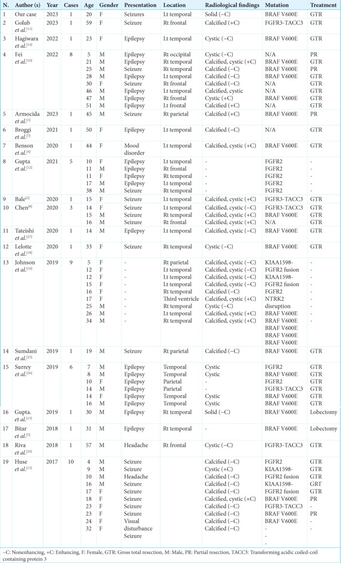
Background: Polymorphous low-grade neuroepithelial tumor of the young (PLNTY) is a newly recognized entity first described in 2017. This article reports the clinical, radiological, histological, and molecular characteristics of PLNTY diagnosed in a young female.
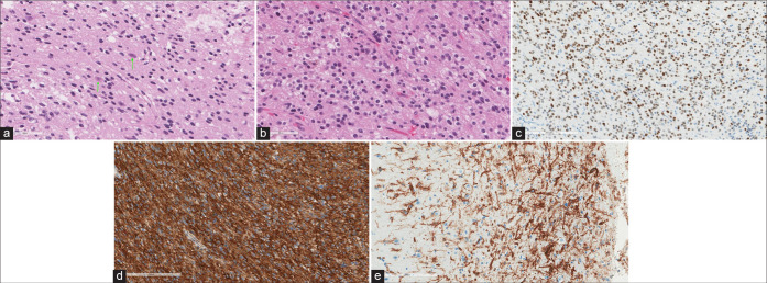
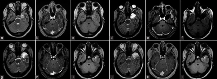
Case description: A 20-year-old female, not known to have any medical illness, presented to the emergency department with speech arrest that progressed to generalized tonic-clonic seizures. Brain magnetic resonance imaging showed a T2 hyperintense nonenhancing cortical-based heterogeneous mass in the left medial temporal lobe and adjacent inferior temporal gyrus measuring 2.7 × 2 × 2.5 cm with no susceptibility signal or diffusion restriction. Electroencephalogram (EEG) showed left temporal epileptiform discharges. She underwent left frontotemporal craniotomy and tumor resection. The light microscopic examination of the tumor revealed a low-grade infiltrative neoplasm. There was a classical appearance of round cells with perinuclear halo, immunopositive for olig-2, glial fibrillary acidic protein (GFAP), and cluster of differentiation-34. There was B-Raf protooncogene, serine/threonine kinase (BRAF) c.1799T>A (p.V600E) point mutation and absence of isocitrate dehydrogenase (IDH) 1 and 2 hotspot mutations. The overall findings were diagnostic of PLNTY.
Conclusion: This article reports an additional case of PLNTY, a newly defined central nervous system tumor entity, describing its clinical, radiological, histological, and molecular features.

 求助内容:
求助内容: 应助结果提醒方式:


