David Holthaus, Nina Hedemann, Sophia Theresa Geweniger, Huy Duc Le, Ibrahim Alkatout, Mirjana Kessler, Thomas F Meyer
{"title":"建立和维持人输卵管上皮三维类器官培养。","authors":"David Holthaus, Nina Hedemann, Sophia Theresa Geweniger, Huy Duc Le, Ibrahim Alkatout, Mirjana Kessler, Thomas F Meyer","doi":"10.21769/BioProtoc.5412","DOIUrl":null,"url":null,"abstract":"<p><p>The female reproductive tract is comprised of different regions, each with distinctive physiological characteristics. One of them is the fallopian tubes, which are vital for human reproductive health and success. The ability to model their function and physiology is of utmost importance. So far, in vitro models have been based on a few immortalized or cancer cell lines derived from fallopian tube cells that lacked differentiated, specialized cell types and did not allow for the study of cancer initiation due to their implicit biases. Organoids, in contrast, overcome these limitations and provide an advanced, three-dimensional system for the study of healthy fallopian tube physiology and pathology. Fallopian tube organoids are comprised of epithelial progenitors that can be enriched using chemical or hormonal treatment into the different cell types that are found in the in vivo tissue, namely detyrosinated-tubulin-positive ciliated cells or paired-box protein 8 (PAX8)-positive secretory cells. This protocol provides a step-by-step guide for the establishment and maintenance of a long-term culture of organoids from healthy human fallopian tube tissue. The organoid model described here closely mimics the in vivo physiology and anatomy of human fallopian tube epithelium and provides a comprehensive basis for future studies on its underlying molecular characteristics and possible pathology. Key features • Provides a step-by-step guide for the establishment of long-term fallopian tube organoid cultures. • Allows for rapid extension of fallopian tube epithelial progenitor cells with a yield of up to 1 × 10<sup>8</sup> cells within 3 weeks of isolation. • Fallopian tube organoids closely mimic healthy physiology, being comprised of multiple different cell types, like detyrosinated-tubulin-positive ciliated cells or paired-box protein 8 (PAX8)-positive secretory cells. • Further enrichment of secretory cells by hormonal treatment and ciliated cells by chemical treatment is possible.</p>","PeriodicalId":93907,"journal":{"name":"Bio-protocol","volume":"15 16","pages":"e5412"},"PeriodicalIF":1.1000,"publicationDate":"2025-08-20","publicationTypes":"Journal Article","fieldsOfStudy":null,"isOpenAccess":false,"openAccessPdf":"https://www.ncbi.nlm.nih.gov/pmc/articles/PMC12378425/pdf/","citationCount":"0","resultStr":"{\"title\":\"Establishing and Maintaining 3D Organoid Cultures From Human Fallopian Tube Epithelium.\",\"authors\":\"David Holthaus, Nina Hedemann, Sophia Theresa Geweniger, Huy Duc Le, Ibrahim Alkatout, Mirjana Kessler, Thomas F Meyer\",\"doi\":\"10.21769/BioProtoc.5412\",\"DOIUrl\":null,\"url\":null,\"abstract\":\"<p><p>The female reproductive tract is comprised of different regions, each with distinctive physiological characteristics. One of them is the fallopian tubes, which are vital for human reproductive health and success. The ability to model their function and physiology is of utmost importance. So far, in vitro models have been based on a few immortalized or cancer cell lines derived from fallopian tube cells that lacked differentiated, specialized cell types and did not allow for the study of cancer initiation due to their implicit biases. Organoids, in contrast, overcome these limitations and provide an advanced, three-dimensional system for the study of healthy fallopian tube physiology and pathology. Fallopian tube organoids are comprised of epithelial progenitors that can be enriched using chemical or hormonal treatment into the different cell types that are found in the in vivo tissue, namely detyrosinated-tubulin-positive ciliated cells or paired-box protein 8 (PAX8)-positive secretory cells. This protocol provides a step-by-step guide for the establishment and maintenance of a long-term culture of organoids from healthy human fallopian tube tissue. The organoid model described here closely mimics the in vivo physiology and anatomy of human fallopian tube epithelium and provides a comprehensive basis for future studies on its underlying molecular characteristics and possible pathology. Key features • Provides a step-by-step guide for the establishment of long-term fallopian tube organoid cultures. • Allows for rapid extension of fallopian tube epithelial progenitor cells with a yield of up to 1 × 10<sup>8</sup> cells within 3 weeks of isolation. • Fallopian tube organoids closely mimic healthy physiology, being comprised of multiple different cell types, like detyrosinated-tubulin-positive ciliated cells or paired-box protein 8 (PAX8)-positive secretory cells. • Further enrichment of secretory cells by hormonal treatment and ciliated cells by chemical treatment is possible.</p>\",\"PeriodicalId\":93907,\"journal\":{\"name\":\"Bio-protocol\",\"volume\":\"15 16\",\"pages\":\"e5412\"},\"PeriodicalIF\":1.1000,\"publicationDate\":\"2025-08-20\",\"publicationTypes\":\"Journal Article\",\"fieldsOfStudy\":null,\"isOpenAccess\":false,\"openAccessPdf\":\"https://www.ncbi.nlm.nih.gov/pmc/articles/PMC12378425/pdf/\",\"citationCount\":\"0\",\"resultStr\":null,\"platform\":\"Semanticscholar\",\"paperid\":null,\"PeriodicalName\":\"Bio-protocol\",\"FirstCategoryId\":\"1085\",\"ListUrlMain\":\"https://doi.org/10.21769/BioProtoc.5412\",\"RegionNum\":0,\"RegionCategory\":null,\"ArticlePicture\":[],\"TitleCN\":null,\"AbstractTextCN\":null,\"PMCID\":null,\"EPubDate\":\"\",\"PubModel\":\"\",\"JCR\":\"Q3\",\"JCRName\":\"BIOLOGY\",\"Score\":null,\"Total\":0}","platform":"Semanticscholar","paperid":null,"PeriodicalName":"Bio-protocol","FirstCategoryId":"1085","ListUrlMain":"https://doi.org/10.21769/BioProtoc.5412","RegionNum":0,"RegionCategory":null,"ArticlePicture":[],"TitleCN":null,"AbstractTextCN":null,"PMCID":null,"EPubDate":"","PubModel":"","JCR":"Q3","JCRName":"BIOLOGY","Score":null,"Total":0}
Establishing and Maintaining 3D Organoid Cultures From Human Fallopian Tube Epithelium.
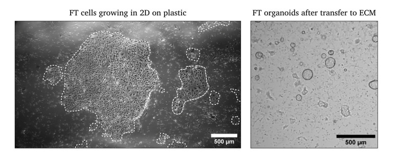
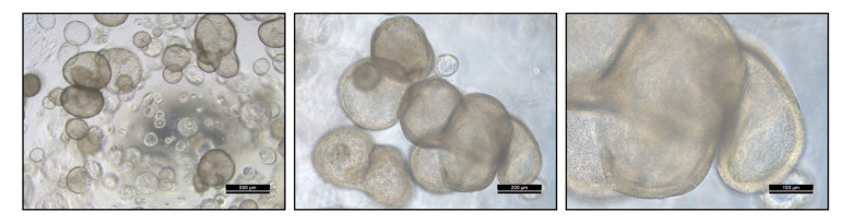
The female reproductive tract is comprised of different regions, each with distinctive physiological characteristics. One of them is the fallopian tubes, which are vital for human reproductive health and success. The ability to model their function and physiology is of utmost importance. So far, in vitro models have been based on a few immortalized or cancer cell lines derived from fallopian tube cells that lacked differentiated, specialized cell types and did not allow for the study of cancer initiation due to their implicit biases. Organoids, in contrast, overcome these limitations and provide an advanced, three-dimensional system for the study of healthy fallopian tube physiology and pathology. Fallopian tube organoids are comprised of epithelial progenitors that can be enriched using chemical or hormonal treatment into the different cell types that are found in the in vivo tissue, namely detyrosinated-tubulin-positive ciliated cells or paired-box protein 8 (PAX8)-positive secretory cells. This protocol provides a step-by-step guide for the establishment and maintenance of a long-term culture of organoids from healthy human fallopian tube tissue. The organoid model described here closely mimics the in vivo physiology and anatomy of human fallopian tube epithelium and provides a comprehensive basis for future studies on its underlying molecular characteristics and possible pathology. Key features • Provides a step-by-step guide for the establishment of long-term fallopian tube organoid cultures. • Allows for rapid extension of fallopian tube epithelial progenitor cells with a yield of up to 1 × 10⁸ cells within 3 weeks of isolation. • Fallopian tube organoids closely mimic healthy physiology, being comprised of multiple different cell types, like detyrosinated-tubulin-positive ciliated cells or paired-box protein 8 (PAX8)-positive secretory cells. • Further enrichment of secretory cells by hormonal treatment and ciliated cells by chemical treatment is possible.

 求助内容:
求助内容: 应助结果提醒方式:


