Yao Chen, Savannah M Decker, Petr Bruza, David J Gladstone, Lesley A Jarvis, Brian W Pogue, Kimberley S Samkoe, Rongxiao Zhang
{"title":"切伦科夫成像生物形态学特征验证乳房放疗中可变形组织移位的患者定位。","authors":"Yao Chen, Savannah M Decker, Petr Bruza, David J Gladstone, Lesley A Jarvis, Brian W Pogue, Kimberley S Samkoe, Rongxiao Zhang","doi":"","DOIUrl":null,"url":null,"abstract":"<p><strong>Purpose: </strong>Accurate patient positioning is crucial for precise radiation therapy dose delivery, as errors in positioning can profoundly influence treatment outcomes. This study introduces a novel application for loco-regional tissue deformation tracking via Cherenkov image analysis during fractionated breast cancer radiation therapy. The primary objective of this research was to develop and test an algorithmic method for Cherenkov-based position accuracy quantification, particularly for loco-regional deformations, which do not have an ideal method for quantification during radiation therapy.</p><p><strong>Methods and materials: </strong>Bio-morphological features in the Cherenkov images, such as vessels, were segmented. A rigid/nonrigid combined registration technique was employed to pinpoint both inter- and intrafractional positioning variations. The methodology was tested on an anthropomorphic chest phantom experiment via shifting a treatment couch with known distances and inducing respiratory motion to simulate interfraction setup uncertainties and intrafraction motions, respectively. It was then applied to a data set of fractionated whole breast radiation therapy human imaging (n = 10 patients).</p><p><strong>Results: </strong>The methodology provided quantified positioning variations comprising 2 components: a global shift determined through rigid registration and a 2-dimensional variation map illustrating loco-regional tissue deformation quantified via nonrigid registration. Controlled phantom testing yielded an average accuracy of 0.83 mm for couch translations up to 20 mm in various directions. Analysis of clinical Cherenkov imaging data from 10 breast cancer patients compared with their first imaged fraction revealed an interfraction setup variation of 3.7 ± 2.4 mm in the global shift and loco-regional deformation up to 3.3 ± 1.9 mm (95th percentile of all regional deformation).</p><p><strong>Conclusions: </strong>This study introduces the use of Cherenkov visualized bio-morphological features to quantify the global and local variations in patient positioning based on rigid and nonrigid registrations. This new approach demonstrates the feasibility of providing quantitative guidance for inter/intrafraction positioning, particularly for the loco-regional deformations that have been unappreciated in current practice with conventional imaging techniques.</p>","PeriodicalId":93888,"journal":{"name":"ArXiv","volume":" ","pages":""},"PeriodicalIF":0.0000,"publicationDate":"2025-08-19","publicationTypes":"Journal Article","fieldsOfStudy":null,"isOpenAccess":false,"openAccessPdf":"https://www.ncbi.nlm.nih.gov/pmc/articles/PMC12393243/pdf/","citationCount":"0","resultStr":"{\"title\":\"Cherenkov Imaged Bio-Morphological Features Verify Patient Positioning With Deformable Tissue Translocation in Breast Radiation Therapy.\",\"authors\":\"Yao Chen, Savannah M Decker, Petr Bruza, David J Gladstone, Lesley A Jarvis, Brian W Pogue, Kimberley S Samkoe, Rongxiao Zhang\",\"doi\":\"\",\"DOIUrl\":null,\"url\":null,\"abstract\":\"<p><strong>Purpose: </strong>Accurate patient positioning is crucial for precise radiation therapy dose delivery, as errors in positioning can profoundly influence treatment outcomes. This study introduces a novel application for loco-regional tissue deformation tracking via Cherenkov image analysis during fractionated breast cancer radiation therapy. The primary objective of this research was to develop and test an algorithmic method for Cherenkov-based position accuracy quantification, particularly for loco-regional deformations, which do not have an ideal method for quantification during radiation therapy.</p><p><strong>Methods and materials: </strong>Bio-morphological features in the Cherenkov images, such as vessels, were segmented. A rigid/nonrigid combined registration technique was employed to pinpoint both inter- and intrafractional positioning variations. The methodology was tested on an anthropomorphic chest phantom experiment via shifting a treatment couch with known distances and inducing respiratory motion to simulate interfraction setup uncertainties and intrafraction motions, respectively. It was then applied to a data set of fractionated whole breast radiation therapy human imaging (n = 10 patients).</p><p><strong>Results: </strong>The methodology provided quantified positioning variations comprising 2 components: a global shift determined through rigid registration and a 2-dimensional variation map illustrating loco-regional tissue deformation quantified via nonrigid registration. Controlled phantom testing yielded an average accuracy of 0.83 mm for couch translations up to 20 mm in various directions. Analysis of clinical Cherenkov imaging data from 10 breast cancer patients compared with their first imaged fraction revealed an interfraction setup variation of 3.7 ± 2.4 mm in the global shift and loco-regional deformation up to 3.3 ± 1.9 mm (95th percentile of all regional deformation).</p><p><strong>Conclusions: </strong>This study introduces the use of Cherenkov visualized bio-morphological features to quantify the global and local variations in patient positioning based on rigid and nonrigid registrations. This new approach demonstrates the feasibility of providing quantitative guidance for inter/intrafraction positioning, particularly for the loco-regional deformations that have been unappreciated in current practice with conventional imaging techniques.</p>\",\"PeriodicalId\":93888,\"journal\":{\"name\":\"ArXiv\",\"volume\":\" \",\"pages\":\"\"},\"PeriodicalIF\":0.0000,\"publicationDate\":\"2025-08-19\",\"publicationTypes\":\"Journal Article\",\"fieldsOfStudy\":null,\"isOpenAccess\":false,\"openAccessPdf\":\"https://www.ncbi.nlm.nih.gov/pmc/articles/PMC12393243/pdf/\",\"citationCount\":\"0\",\"resultStr\":null,\"platform\":\"Semanticscholar\",\"paperid\":null,\"PeriodicalName\":\"ArXiv\",\"FirstCategoryId\":\"1085\",\"ListUrlMain\":\"\",\"RegionNum\":0,\"RegionCategory\":null,\"ArticlePicture\":[],\"TitleCN\":null,\"AbstractTextCN\":null,\"PMCID\":null,\"EPubDate\":\"\",\"PubModel\":\"\",\"JCR\":\"\",\"JCRName\":\"\",\"Score\":null,\"Total\":0}","platform":"Semanticscholar","paperid":null,"PeriodicalName":"ArXiv","FirstCategoryId":"1085","ListUrlMain":"","RegionNum":0,"RegionCategory":null,"ArticlePicture":[],"TitleCN":null,"AbstractTextCN":null,"PMCID":null,"EPubDate":"","PubModel":"","JCR":"","JCRName":"","Score":null,"Total":0}
Cherenkov Imaged Bio-Morphological Features Verify Patient Positioning With Deformable Tissue Translocation in Breast Radiation Therapy.
Purpose: Accurate patient positioning is crucial for precise radiation therapy dose delivery, as errors in positioning can profoundly influence treatment outcomes. This study introduces a novel application for loco-regional tissue deformation tracking via Cherenkov image analysis during fractionated breast cancer radiation therapy. The primary objective of this research was to develop and test an algorithmic method for Cherenkov-based position accuracy quantification, particularly for loco-regional deformations, which do not have an ideal method for quantification during radiation therapy.
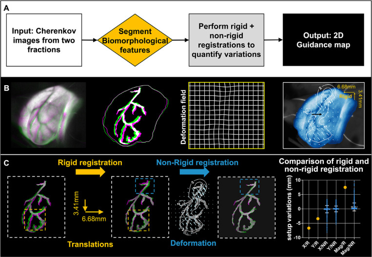
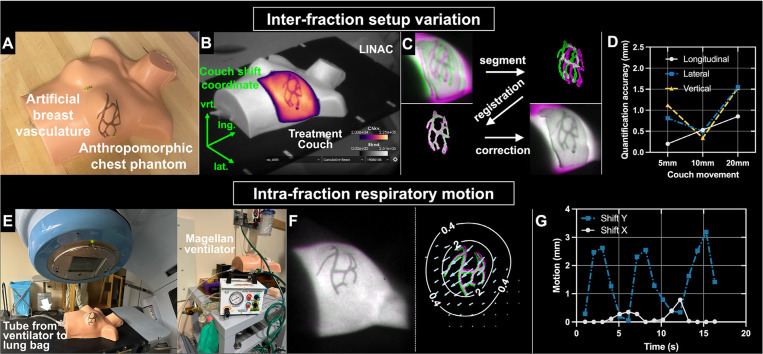
Methods and materials: Bio-morphological features in the Cherenkov images, such as vessels, were segmented. A rigid/nonrigid combined registration technique was employed to pinpoint both inter- and intrafractional positioning variations. The methodology was tested on an anthropomorphic chest phantom experiment via shifting a treatment couch with known distances and inducing respiratory motion to simulate interfraction setup uncertainties and intrafraction motions, respectively. It was then applied to a data set of fractionated whole breast radiation therapy human imaging (n = 10 patients).
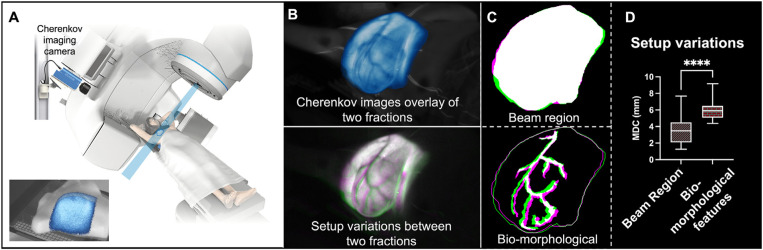
Results: The methodology provided quantified positioning variations comprising 2 components: a global shift determined through rigid registration and a 2-dimensional variation map illustrating loco-regional tissue deformation quantified via nonrigid registration. Controlled phantom testing yielded an average accuracy of 0.83 mm for couch translations up to 20 mm in various directions. Analysis of clinical Cherenkov imaging data from 10 breast cancer patients compared with their first imaged fraction revealed an interfraction setup variation of 3.7 ± 2.4 mm in the global shift and loco-regional deformation up to 3.3 ± 1.9 mm (95th percentile of all regional deformation).
Conclusions: This study introduces the use of Cherenkov visualized bio-morphological features to quantify the global and local variations in patient positioning based on rigid and nonrigid registrations. This new approach demonstrates the feasibility of providing quantitative guidance for inter/intrafraction positioning, particularly for the loco-regional deformations that have been unappreciated in current practice with conventional imaging techniques.

 求助内容:
求助内容: 应助结果提醒方式:


