Evangelia Ntikoudi, Thomas Karagkounis, Konstantinos S Mylonas, Stylianos Kykalos, Dimitrios Schizas, Ioannis N Vamvakaris, Ekaterini Politi, Michail V Karamouzis, Stamatios Theocharis
{"title":"PROX1在切除的非小细胞肺癌中的表达:免疫组织化学特征和临床病理相关性。","authors":"Evangelia Ntikoudi, Thomas Karagkounis, Konstantinos S Mylonas, Stylianos Kykalos, Dimitrios Schizas, Ioannis N Vamvakaris, Ekaterini Politi, Michail V Karamouzis, Stamatios Theocharis","doi":"10.3390/medsci13030140","DOIUrl":null,"url":null,"abstract":"<p><p><b>Background/Objectives:</b> PROX1 (prospero homeobox 1) is a transcription factor involved in lymphangiogenesis and cellular differentiation. Its role in cancer biology appears to be highly context-dependent, with it exhibiting both tumor-promoting and -suppressive functions across various malignancies. Nonetheless, the clinical significance of PROX1 expression in non-small cell lung cancer (NSCLC) remains poorly elucidated. The objective of this study is to evaluate the immunohistochemical expression of PROX1 in NSCLC, specifically in the adenocarcinoma and squamous cell carcinoma subtypes, and to assess its correlation with clinicopathologic features and overall survival (OS). <b>Methods:</b> This retrospective study included surgically resected specimens from 121 patients with histologically confirmed NSCLC. PROX1 expression was assessed via immunohistochemistry on formalin-fixed, paraffin-embedded specimens. Staining intensity (graded 0- National and Kapodistrian University of Athens 3) and the percentage of positive tumor cells were recorded. Correlations with histological subtype, tumor characteristics, and OS were analyzed using chi-square tests, one-way ANOVA, and Kaplan-Meier survival analysis with log-rank testing. <b>Results:</b> Low PROX1 intensity (level 1) was significantly associated with P63 positivity (<i>p</i> = 0.028), while high PROX1 intensity (level 3) correlated with nodal metastasis to station 3 (S3+) (<i>p</i> = 0.025). Additionally, alveolar-pattern adenocarcinomas exhibited intermediate PROX1 expression (26-50%) (<i>p</i> = 0.010). Although PROX1 positivity did not differ among mucinous and non-mucinous adenocarcinomas (<i>p</i> = 0.152), its distribution across defined expression subgroups was statistically significant (<i>p</i> = 0.002). Tumors with low PROX1 expression (0-24%) were associated with a larger maximum tumor diameter (<i>p</i> = 0.026). PROX1 expression was not independently associated with OS (<i>p</i> > 0.05). Factors significantly associated with improved survival included an age < 50 years, female sex, the absence of necrosis, fewer than 10 positive lymph nodes, a lymph node ratio < 0.5, and the absence of extensive nodal involvement in stations 5, 10, 11, and 12. <b>Conclusions:</b> Although PROX1 expression is variably associated with specific histologic subtypes and nodal metastases in NSCLC, it does not independently predict overall survival. Its expression patterns suggest a potential role in tumor differentiation and lymphatic spread. Further mechanistic and immunologic studies are warranted to elucidate the functional significance of PROX1 in lung cancer biology.</p>","PeriodicalId":74152,"journal":{"name":"Medical sciences (Basel, Switzerland)","volume":"13 3","pages":""},"PeriodicalIF":4.4000,"publicationDate":"2025-08-17","publicationTypes":"Journal Article","fieldsOfStudy":null,"isOpenAccess":false,"openAccessPdf":"https://www.ncbi.nlm.nih.gov/pmc/articles/PMC12372085/pdf/","citationCount":"0","resultStr":"{\"title\":\"PROX1 Expression in Resected Non-Small Cell Lung Cancer: Immunohistochemical Profile and Clinicopathological Correlates.\",\"authors\":\"Evangelia Ntikoudi, Thomas Karagkounis, Konstantinos S Mylonas, Stylianos Kykalos, Dimitrios Schizas, Ioannis N Vamvakaris, Ekaterini Politi, Michail V Karamouzis, Stamatios Theocharis\",\"doi\":\"10.3390/medsci13030140\",\"DOIUrl\":null,\"url\":null,\"abstract\":\"<p><p><b>Background/Objectives:</b> PROX1 (prospero homeobox 1) is a transcription factor involved in lymphangiogenesis and cellular differentiation. Its role in cancer biology appears to be highly context-dependent, with it exhibiting both tumor-promoting and -suppressive functions across various malignancies. Nonetheless, the clinical significance of PROX1 expression in non-small cell lung cancer (NSCLC) remains poorly elucidated. The objective of this study is to evaluate the immunohistochemical expression of PROX1 in NSCLC, specifically in the adenocarcinoma and squamous cell carcinoma subtypes, and to assess its correlation with clinicopathologic features and overall survival (OS). <b>Methods:</b> This retrospective study included surgically resected specimens from 121 patients with histologically confirmed NSCLC. PROX1 expression was assessed via immunohistochemistry on formalin-fixed, paraffin-embedded specimens. Staining intensity (graded 0- National and Kapodistrian University of Athens 3) and the percentage of positive tumor cells were recorded. Correlations with histological subtype, tumor characteristics, and OS were analyzed using chi-square tests, one-way ANOVA, and Kaplan-Meier survival analysis with log-rank testing. <b>Results:</b> Low PROX1 intensity (level 1) was significantly associated with P63 positivity (<i>p</i> = 0.028), while high PROX1 intensity (level 3) correlated with nodal metastasis to station 3 (S3+) (<i>p</i> = 0.025). Additionally, alveolar-pattern adenocarcinomas exhibited intermediate PROX1 expression (26-50%) (<i>p</i> = 0.010). Although PROX1 positivity did not differ among mucinous and non-mucinous adenocarcinomas (<i>p</i> = 0.152), its distribution across defined expression subgroups was statistically significant (<i>p</i> = 0.002). Tumors with low PROX1 expression (0-24%) were associated with a larger maximum tumor diameter (<i>p</i> = 0.026). PROX1 expression was not independently associated with OS (<i>p</i> > 0.05). Factors significantly associated with improved survival included an age < 50 years, female sex, the absence of necrosis, fewer than 10 positive lymph nodes, a lymph node ratio < 0.5, and the absence of extensive nodal involvement in stations 5, 10, 11, and 12. <b>Conclusions:</b> Although PROX1 expression is variably associated with specific histologic subtypes and nodal metastases in NSCLC, it does not independently predict overall survival. Its expression patterns suggest a potential role in tumor differentiation and lymphatic spread. Further mechanistic and immunologic studies are warranted to elucidate the functional significance of PROX1 in lung cancer biology.</p>\",\"PeriodicalId\":74152,\"journal\":{\"name\":\"Medical sciences (Basel, Switzerland)\",\"volume\":\"13 3\",\"pages\":\"\"},\"PeriodicalIF\":4.4000,\"publicationDate\":\"2025-08-17\",\"publicationTypes\":\"Journal Article\",\"fieldsOfStudy\":null,\"isOpenAccess\":false,\"openAccessPdf\":\"https://www.ncbi.nlm.nih.gov/pmc/articles/PMC12372085/pdf/\",\"citationCount\":\"0\",\"resultStr\":null,\"platform\":\"Semanticscholar\",\"paperid\":null,\"PeriodicalName\":\"Medical sciences (Basel, Switzerland)\",\"FirstCategoryId\":\"1085\",\"ListUrlMain\":\"https://doi.org/10.3390/medsci13030140\",\"RegionNum\":0,\"RegionCategory\":null,\"ArticlePicture\":[],\"TitleCN\":null,\"AbstractTextCN\":null,\"PMCID\":null,\"EPubDate\":\"\",\"PubModel\":\"\",\"JCR\":\"Q1\",\"JCRName\":\"Medicine\",\"Score\":null,\"Total\":0}","platform":"Semanticscholar","paperid":null,"PeriodicalName":"Medical sciences (Basel, Switzerland)","FirstCategoryId":"1085","ListUrlMain":"https://doi.org/10.3390/medsci13030140","RegionNum":0,"RegionCategory":null,"ArticlePicture":[],"TitleCN":null,"AbstractTextCN":null,"PMCID":null,"EPubDate":"","PubModel":"","JCR":"Q1","JCRName":"Medicine","Score":null,"Total":0}
引用次数: 0
摘要
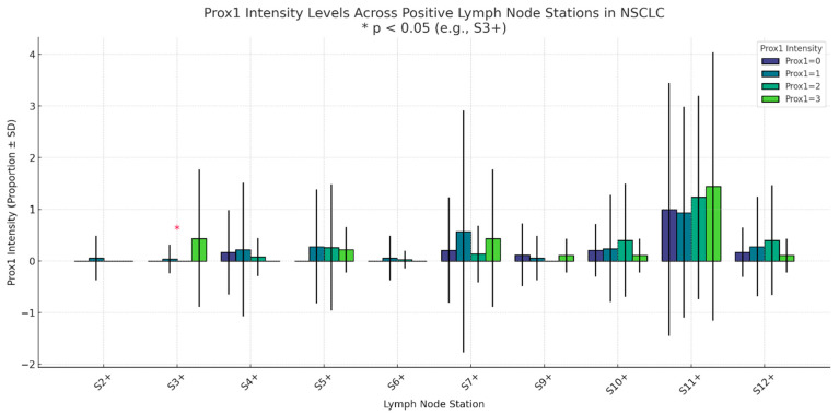
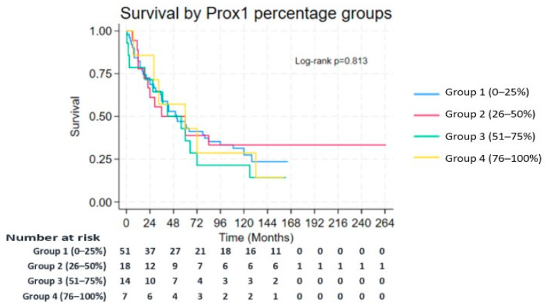
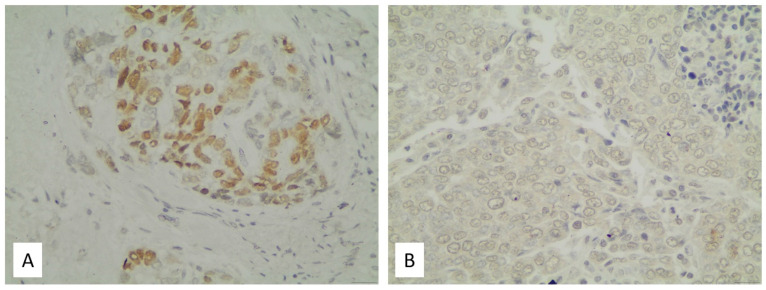
背景/目的:PROX1 (prospero homeobox 1)是一种参与淋巴管生成和细胞分化的转录因子。它在癌症生物学中的作用似乎是高度依赖于环境的,它在各种恶性肿瘤中都表现出促进肿瘤和抑制肿瘤的功能。然而,PROX1表达在非小细胞肺癌(NSCLC)中的临床意义尚不清楚。本研究的目的是评估PROX1在非小细胞肺癌,特别是腺癌和鳞状细胞癌亚型中的免疫组织化学表达,并评估其与临床病理特征和总生存期(OS)的相关性。方法:本回顾性研究包括121例经组织学证实的非小细胞肺癌手术切除标本。免疫组织化学检测福尔马林固定石蜡包埋标本中PROX1的表达。记录染色强度(分级0- National and Kapodistrian University of Athens 3)和阳性肿瘤细胞百分比。采用卡方检验、单因素方差分析和Kaplan-Meier生存分析(log-rank检验)分析与组织学亚型、肿瘤特征和OS的相关性。结果:低PROX1水平(1级)与P63阳性相关(p = 0.028),高PROX1水平(3级)与淋巴结转移至3站(S3+)相关(p = 0.025)。此外,肺泡型腺癌显示PROX1的中等表达(26-50%)(p = 0.010)。虽然PROX1阳性在黏液性腺癌和非黏液性腺癌中没有差异(p = 0.152),但其在定义表达亚组中的分布具有统计学意义(p = 0.002)。低PROX1表达的肿瘤(0-24%)与最大肿瘤直径较大相关(p = 0.026)。PROX1表达与OS无独立相关性(p < 0.05)。与生存率显著相关的因素包括年龄< 50岁,女性,无坏死,少于10个阳性淋巴结,淋巴结比例< 0.5,以及在第5、10、11和12站没有广泛的淋巴结累及。结论:尽管PROX1表达与非小细胞肺癌的特定组织学亚型和淋巴结转移有不同的相关性,但它并不能独立预测总生存率。其表达模式提示其在肿瘤分化和淋巴扩散中有潜在作用。需要进一步的机制和免疫学研究来阐明PROX1在肺癌生物学中的功能意义。
PROX1 Expression in Resected Non-Small Cell Lung Cancer: Immunohistochemical Profile and Clinicopathological Correlates.
Background/Objectives: PROX1 (prospero homeobox 1) is a transcription factor involved in lymphangiogenesis and cellular differentiation. Its role in cancer biology appears to be highly context-dependent, with it exhibiting both tumor-promoting and -suppressive functions across various malignancies. Nonetheless, the clinical significance of PROX1 expression in non-small cell lung cancer (NSCLC) remains poorly elucidated. The objective of this study is to evaluate the immunohistochemical expression of PROX1 in NSCLC, specifically in the adenocarcinoma and squamous cell carcinoma subtypes, and to assess its correlation with clinicopathologic features and overall survival (OS). Methods: This retrospective study included surgically resected specimens from 121 patients with histologically confirmed NSCLC. PROX1 expression was assessed via immunohistochemistry on formalin-fixed, paraffin-embedded specimens. Staining intensity (graded 0- National and Kapodistrian University of Athens 3) and the percentage of positive tumor cells were recorded. Correlations with histological subtype, tumor characteristics, and OS were analyzed using chi-square tests, one-way ANOVA, and Kaplan-Meier survival analysis with log-rank testing. Results: Low PROX1 intensity (level 1) was significantly associated with P63 positivity (p = 0.028), while high PROX1 intensity (level 3) correlated with nodal metastasis to station 3 (S3+) (p = 0.025). Additionally, alveolar-pattern adenocarcinomas exhibited intermediate PROX1 expression (26-50%) (p = 0.010). Although PROX1 positivity did not differ among mucinous and non-mucinous adenocarcinomas (p = 0.152), its distribution across defined expression subgroups was statistically significant (p = 0.002). Tumors with low PROX1 expression (0-24%) were associated with a larger maximum tumor diameter (p = 0.026). PROX1 expression was not independently associated with OS (p > 0.05). Factors significantly associated with improved survival included an age < 50 years, female sex, the absence of necrosis, fewer than 10 positive lymph nodes, a lymph node ratio < 0.5, and the absence of extensive nodal involvement in stations 5, 10, 11, and 12. Conclusions: Although PROX1 expression is variably associated with specific histologic subtypes and nodal metastases in NSCLC, it does not independently predict overall survival. Its expression patterns suggest a potential role in tumor differentiation and lymphatic spread. Further mechanistic and immunologic studies are warranted to elucidate the functional significance of PROX1 in lung cancer biology.

 求助内容:
求助内容: 应助结果提醒方式:


