Samuel Madureira Silva, Frédéric Chalmel, Andrea Errico, Katerina Papageorgiou, Guillaume Richer, Edith Chan Sock Peng, Antoine D Rolland, Kelly Tilleman, Guy T'Sjoen, Ilaria Dando, Tamara Vanhaecke, Ellen Goossens, Yoni Baert
{"title":"从变性女性睾丸组织中提取的细胞型和产生睾酮的类器官体外模型。","authors":"Samuel Madureira Silva, Frédéric Chalmel, Andrea Errico, Katerina Papageorgiou, Guillaume Richer, Edith Chan Sock Peng, Antoine D Rolland, Kelly Tilleman, Guy T'Sjoen, Ilaria Dando, Tamara Vanhaecke, Ellen Goossens, Yoni Baert","doi":"10.1093/hropen/hoaf043","DOIUrl":null,"url":null,"abstract":"<p><strong>Study question: </strong>Can testicular tissue from trans women (trans tissue) be used to create human testicular organoids?</p><p><strong>Summary answer: </strong>Testosterone-producing and cytotypic human testicular organoids with bicompartmental architecture can be successfully generated from trans tissue.</p><p><strong>What is known already: </strong>Testicular organoids are a promising tool for studying testicular function and the effects of toxicants. Immature testicular cells are currently the most efficient at forming organoids that closely recapitulate seminiferous tubule-like architecture and functions. However, the scarcity of immature human testicular tissue limits its use in high-throughput applications. Conversely, trans tissue is abundantly available and characterized by an immature phenotype.</p><p><strong>Study design size duration: </strong>Trans tissue-derived organoids (trans organoids) were histologically and androgenically compared to reference organoids derived from immature (prepubertal and pubertal) and adult cisgender testicular tissues. Additionally, long-term testosterone production and gonadotrophic stimulation were assessed in trans organoids. To evaluate their cytotypic and transcriptomic resemblance to reference testicular tissue stages, trans organoids were compared at the gene expression level to prepubertal, pubertal, and adult cisgender tissues, along with their tissue of origin.</p><p><strong>Participants/materials setting methods: </strong>Testicular tissue samples from transgender women, as well as from prepubertal, pubertal, and adult cisgender donors, were used to generate testicular organoids and to compare organoid formation efficiency and testosterone production according to tissue origin. These samples also served as references for transcriptomic comparisons with organoids derived from transgender women's testicular tissue at Day 14 of culture. Testicular organoids were generated and cultured using 3D Petri Dish<sup>®</sup> platforms. Histochemistry and immunofluorescence staining were employed to characterize cellular composition and spatial organization. Testosterone production in culture media was assessed using electrochemiluminescence immunoassays. RNA was extracted and sequenced from organoids derived from transgender women, as well as from tissue samples of all donor groups. Deconvolution and differential gene expression analyses were performed to compare the organoids with testicular tissues across all groups.</p><p><strong>Main results and the role of chance: </strong>Trans organoids form compartmentalized, cytotypic <i>de novo</i> tissues similar to those from pubertal testicular tissue. Additionally, trans organoids exhibit significant testosterone production, sustain this function over extended culture periods, and respond to gonadotrophic stimulation. Deconvolved bulk RNAseq data indicate that cell population proportions within these organoids are close to those in prepubertal and pubertal testicular tissues. Gene expression clusters trans organoids alongside prepubertal and trans tissues. Functional analysis reveals that trans organoids share with prepubertal, pubertal, and trans tissues varied cellular processes. Factors such as the duration of hormone therapy, the expression of anti-Müllerian hormone-an immaturity marker-within the tubules, and the proportion of peritubular myoid cells in the donor tissue were found to predict the success of trans organoid formation.</p><p><strong>Large scale data: </strong>The bulk RNA-seq raw and preprocessed data are stored under restricted access in the Vrije Universiteit Brussel (VUB) Institutional Data Repository (VUB/IVTD/1/000001) due to participant privacy concerns. Access to the data will be considered by contacting Prof. Yoni Baert (yoni.baert@vub.be).</p><p><strong>Limitations reasons for caution: </strong>Hormonal data from trans women donors were not acquired in a convenient manner for this study. Deconvolution data allow only cell proportions to be compared, not absolute numbers.</p><p><strong>Wider implications of the findings: </strong>This study highlights the potential of trans organoids as a novel and ethically sustainable human-based model for male reproductive health research, reproductive toxicology, and endocrine disruption studies. While trans tissue is a valuable replacement for immature tissue, further research should focus on optimizing organoid architecture, evaluating their utility in reprotoxicity testing, and promoting germ cell differentiation.</p><p><strong>Study funding/competing interests: </strong>This study was conducted with financial support from the VUB Research Council (OZR4004) to S.M.S., the Scientific Research Foundation-Flanders (G026223N) and the Scientific Fund Willy Gepts to Y.B., the Strategic Research Program 89 from the VUB to E.G., and the Mireille Aerens Chair to T.V. The authors declare no conflict of interest.</p>","PeriodicalId":73264,"journal":{"name":"Human reproduction open","volume":"2025 3","pages":"hoaf043"},"PeriodicalIF":11.1000,"publicationDate":"2025-08-20","publicationTypes":"Journal Article","fieldsOfStudy":null,"isOpenAccess":false,"openAccessPdf":"https://www.ncbi.nlm.nih.gov/pmc/articles/PMC12396852/pdf/","citationCount":"0","resultStr":"{\"title\":\"A new human <i>in vitro</i> model of cytotypic and testosterone-producing organoids derived from testicular tissue of transgender women.\",\"authors\":\"Samuel Madureira Silva, Frédéric Chalmel, Andrea Errico, Katerina Papageorgiou, Guillaume Richer, Edith Chan Sock Peng, Antoine D Rolland, Kelly Tilleman, Guy T'Sjoen, Ilaria Dando, Tamara Vanhaecke, Ellen Goossens, Yoni Baert\",\"doi\":\"10.1093/hropen/hoaf043\",\"DOIUrl\":null,\"url\":null,\"abstract\":\"<p><strong>Study question: </strong>Can testicular tissue from trans women (trans tissue) be used to create human testicular organoids?</p><p><strong>Summary answer: </strong>Testosterone-producing and cytotypic human testicular organoids with bicompartmental architecture can be successfully generated from trans tissue.</p><p><strong>What is known already: </strong>Testicular organoids are a promising tool for studying testicular function and the effects of toxicants. Immature testicular cells are currently the most efficient at forming organoids that closely recapitulate seminiferous tubule-like architecture and functions. However, the scarcity of immature human testicular tissue limits its use in high-throughput applications. Conversely, trans tissue is abundantly available and characterized by an immature phenotype.</p><p><strong>Study design size duration: </strong>Trans tissue-derived organoids (trans organoids) were histologically and androgenically compared to reference organoids derived from immature (prepubertal and pubertal) and adult cisgender testicular tissues. Additionally, long-term testosterone production and gonadotrophic stimulation were assessed in trans organoids. To evaluate their cytotypic and transcriptomic resemblance to reference testicular tissue stages, trans organoids were compared at the gene expression level to prepubertal, pubertal, and adult cisgender tissues, along with their tissue of origin.</p><p><strong>Participants/materials setting methods: </strong>Testicular tissue samples from transgender women, as well as from prepubertal, pubertal, and adult cisgender donors, were used to generate testicular organoids and to compare organoid formation efficiency and testosterone production according to tissue origin. These samples also served as references for transcriptomic comparisons with organoids derived from transgender women's testicular tissue at Day 14 of culture. Testicular organoids were generated and cultured using 3D Petri Dish<sup>®</sup> platforms. Histochemistry and immunofluorescence staining were employed to characterize cellular composition and spatial organization. Testosterone production in culture media was assessed using electrochemiluminescence immunoassays. RNA was extracted and sequenced from organoids derived from transgender women, as well as from tissue samples of all donor groups. Deconvolution and differential gene expression analyses were performed to compare the organoids with testicular tissues across all groups.</p><p><strong>Main results and the role of chance: </strong>Trans organoids form compartmentalized, cytotypic <i>de novo</i> tissues similar to those from pubertal testicular tissue. Additionally, trans organoids exhibit significant testosterone production, sustain this function over extended culture periods, and respond to gonadotrophic stimulation. Deconvolved bulk RNAseq data indicate that cell population proportions within these organoids are close to those in prepubertal and pubertal testicular tissues. Gene expression clusters trans organoids alongside prepubertal and trans tissues. Functional analysis reveals that trans organoids share with prepubertal, pubertal, and trans tissues varied cellular processes. Factors such as the duration of hormone therapy, the expression of anti-Müllerian hormone-an immaturity marker-within the tubules, and the proportion of peritubular myoid cells in the donor tissue were found to predict the success of trans organoid formation.</p><p><strong>Large scale data: </strong>The bulk RNA-seq raw and preprocessed data are stored under restricted access in the Vrije Universiteit Brussel (VUB) Institutional Data Repository (VUB/IVTD/1/000001) due to participant privacy concerns. Access to the data will be considered by contacting Prof. Yoni Baert (yoni.baert@vub.be).</p><p><strong>Limitations reasons for caution: </strong>Hormonal data from trans women donors were not acquired in a convenient manner for this study. Deconvolution data allow only cell proportions to be compared, not absolute numbers.</p><p><strong>Wider implications of the findings: </strong>This study highlights the potential of trans organoids as a novel and ethically sustainable human-based model for male reproductive health research, reproductive toxicology, and endocrine disruption studies. While trans tissue is a valuable replacement for immature tissue, further research should focus on optimizing organoid architecture, evaluating their utility in reprotoxicity testing, and promoting germ cell differentiation.</p><p><strong>Study funding/competing interests: </strong>This study was conducted with financial support from the VUB Research Council (OZR4004) to S.M.S., the Scientific Research Foundation-Flanders (G026223N) and the Scientific Fund Willy Gepts to Y.B., the Strategic Research Program 89 from the VUB to E.G., and the Mireille Aerens Chair to T.V. The authors declare no conflict of interest.</p>\",\"PeriodicalId\":73264,\"journal\":{\"name\":\"Human reproduction open\",\"volume\":\"2025 3\",\"pages\":\"hoaf043\"},\"PeriodicalIF\":11.1000,\"publicationDate\":\"2025-08-20\",\"publicationTypes\":\"Journal Article\",\"fieldsOfStudy\":null,\"isOpenAccess\":false,\"openAccessPdf\":\"https://www.ncbi.nlm.nih.gov/pmc/articles/PMC12396852/pdf/\",\"citationCount\":\"0\",\"resultStr\":null,\"platform\":\"Semanticscholar\",\"paperid\":null,\"PeriodicalName\":\"Human reproduction open\",\"FirstCategoryId\":\"1085\",\"ListUrlMain\":\"https://doi.org/10.1093/hropen/hoaf043\",\"RegionNum\":0,\"RegionCategory\":null,\"ArticlePicture\":[],\"TitleCN\":null,\"AbstractTextCN\":null,\"PMCID\":null,\"EPubDate\":\"2025/1/1 0:00:00\",\"PubModel\":\"eCollection\",\"JCR\":\"Q1\",\"JCRName\":\"OBSTETRICS & GYNECOLOGY\",\"Score\":null,\"Total\":0}","platform":"Semanticscholar","paperid":null,"PeriodicalName":"Human reproduction open","FirstCategoryId":"1085","ListUrlMain":"https://doi.org/10.1093/hropen/hoaf043","RegionNum":0,"RegionCategory":null,"ArticlePicture":[],"TitleCN":null,"AbstractTextCN":null,"PMCID":null,"EPubDate":"2025/1/1 0:00:00","PubModel":"eCollection","JCR":"Q1","JCRName":"OBSTETRICS & GYNECOLOGY","Score":null,"Total":0}
引用次数: 0
摘要
研究问题:变性女性的睾丸组织(trans tissue)可以用来制造人类睾丸类器官吗?总结回答:具有双室结构的睾酮分泌和细胞型人类睾丸类器官可以成功地从跨组织中产生。已知情况:睾丸类器官是研究睾丸功能和毒物影响的一个很有前途的工具。目前,未成熟的睾丸细胞在形成类器官方面是最有效的,这些类器官与精管一样的结构和功能非常相似。然而,未成熟人类睾丸组织的稀缺性限制了其在高通量应用中的使用。相反,反式组织大量可用,其特征是不成熟的表型。研究设计规模持续时间:与未成熟(青春期前和青春期)和成年顺性睾丸组织的参考类器官相比,反组织来源的类器官在组织学和雄激素学上进行了比较。此外,长期睾酮分泌和促性腺刺激在反类器官中被评估。为了评估它们的细胞型和转录组学与参考睾丸组织阶段的相似性,我们在基因表达水平上比较了反式类器官与青春期前、青春期和成年顺性组织及其起源组织的差异。参与者/材料设置方法:使用变性女性、青春期前、青春期和成年顺性供体的睾丸组织样本来生成睾丸类器官,并根据组织来源比较类器官的形成效率和睾丸激素的产生。这些样本也可以作为转录组学比较的参考,在培养的第14天与来自跨性别女性睾丸组织的类器官进行比较。使用3D Petri Dish®平台生成和培养睾丸类器官。组织化学和免疫荧光染色表征细胞组成和空间组织。使用电化学发光免疫分析法评估培养基中睾酮的产生。从变性女性的类器官以及所有供体组的组织样本中提取RNA并进行测序。进行反褶积和差异基因表达分析,将所有组的类器官与睾丸组织进行比较。主要结果和突变的作用:反式类器官形成区隔化的细胞型新生组织,类似于青春期睾丸组织。此外,反式类器官表现出显著的睾酮分泌,在较长的培养周期内维持这一功能,并对促性腺刺激有反应。去卷积的大量RNAseq数据表明,这些类器官中的细胞群体比例接近青春期前和青春期睾丸组织中的细胞群体比例。基因表达聚集在青春期前和跨组织旁的类器官。功能分析显示,反式类器官与青春期前、青春期和反式组织共享不同的细胞过程。激素治疗的持续时间、小管内抗勒氏激素(一种不成熟的标志物)的表达以及供体组织中小管周围肌样细胞的比例等因素被发现可以预测转化类器官的成功形成。大规模数据:由于参与者隐私问题,大量RNA-seq原始和预处理数据存储在布鲁塞尔自由大学(VUB)机构数据存储库(VUB/IVTD/1/000001)中,访问受限。请联系Yoni Baert教授(yoni.baert@vub.be)获取数据。注意事项的局限性:本研究没有以方便的方式获得变性女性供体的激素数据。反褶积数据只允许比较细胞比例,而不是绝对数字。研究结果的更广泛意义:这项研究强调了反类器官作为一种新的、道德上可持续的基于人类的男性生殖健康研究、生殖毒理学和内分泌干扰研究模型的潜力。虽然反式组织是一种有价值的未成熟组织替代品,但进一步的研究应集中在优化类器官结构、评估其在生殖毒性测试中的应用以及促进生殖细胞分化方面。研究经费/利益冲突:本研究得到了VUB研究委员会(OZR4004)到s.m.s.,科学研究基金会-弗兰德斯(G026223N)和科学基金Willy Gepts到y.b.,战略研究计划89从VUB到e.g.和Mireille Aerens主席到T.V.的财政支持。作者声明没有利益冲突。
A new human in vitro model of cytotypic and testosterone-producing organoids derived from testicular tissue of transgender women.
Study question: Can testicular tissue from trans women (trans tissue) be used to create human testicular organoids?
Summary answer: Testosterone-producing and cytotypic human testicular organoids with bicompartmental architecture can be successfully generated from trans tissue.
What is known already: Testicular organoids are a promising tool for studying testicular function and the effects of toxicants. Immature testicular cells are currently the most efficient at forming organoids that closely recapitulate seminiferous tubule-like architecture and functions. However, the scarcity of immature human testicular tissue limits its use in high-throughput applications. Conversely, trans tissue is abundantly available and characterized by an immature phenotype.
Study design size duration: Trans tissue-derived organoids (trans organoids) were histologically and androgenically compared to reference organoids derived from immature (prepubertal and pubertal) and adult cisgender testicular tissues. Additionally, long-term testosterone production and gonadotrophic stimulation were assessed in trans organoids. To evaluate their cytotypic and transcriptomic resemblance to reference testicular tissue stages, trans organoids were compared at the gene expression level to prepubertal, pubertal, and adult cisgender tissues, along with their tissue of origin.
Participants/materials setting methods: Testicular tissue samples from transgender women, as well as from prepubertal, pubertal, and adult cisgender donors, were used to generate testicular organoids and to compare organoid formation efficiency and testosterone production according to tissue origin. These samples also served as references for transcriptomic comparisons with organoids derived from transgender women's testicular tissue at Day 14 of culture. Testicular organoids were generated and cultured using 3D Petri Dish® platforms. Histochemistry and immunofluorescence staining were employed to characterize cellular composition and spatial organization. Testosterone production in culture media was assessed using electrochemiluminescence immunoassays. RNA was extracted and sequenced from organoids derived from transgender women, as well as from tissue samples of all donor groups. Deconvolution and differential gene expression analyses were performed to compare the organoids with testicular tissues across all groups.
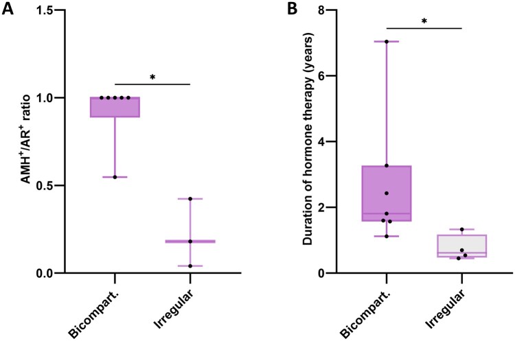
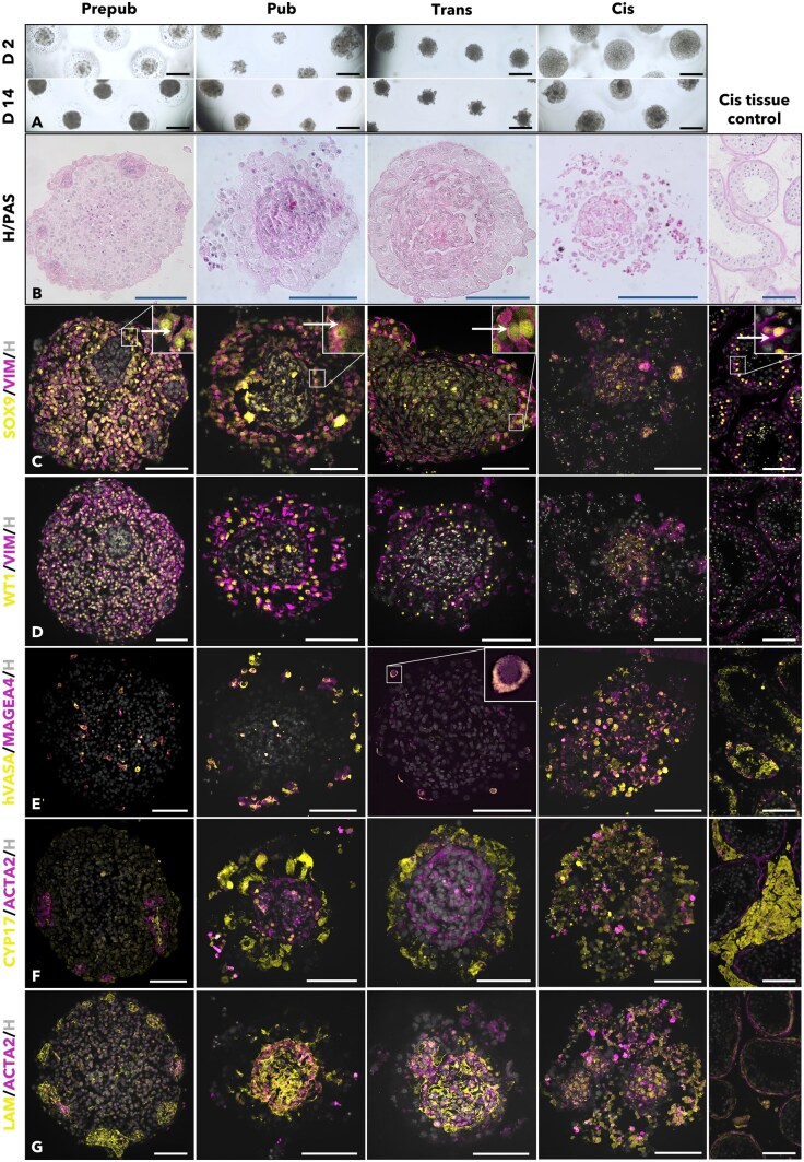
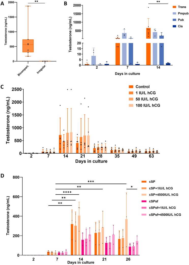
Main results and the role of chance: Trans organoids form compartmentalized, cytotypic de novo tissues similar to those from pubertal testicular tissue. Additionally, trans organoids exhibit significant testosterone production, sustain this function over extended culture periods, and respond to gonadotrophic stimulation. Deconvolved bulk RNAseq data indicate that cell population proportions within these organoids are close to those in prepubertal and pubertal testicular tissues. Gene expression clusters trans organoids alongside prepubertal and trans tissues. Functional analysis reveals that trans organoids share with prepubertal, pubertal, and trans tissues varied cellular processes. Factors such as the duration of hormone therapy, the expression of anti-Müllerian hormone-an immaturity marker-within the tubules, and the proportion of peritubular myoid cells in the donor tissue were found to predict the success of trans organoid formation.
Large scale data: The bulk RNA-seq raw and preprocessed data are stored under restricted access in the Vrije Universiteit Brussel (VUB) Institutional Data Repository (VUB/IVTD/1/000001) due to participant privacy concerns. Access to the data will be considered by contacting Prof. Yoni Baert (yoni.baert@vub.be).
Limitations reasons for caution: Hormonal data from trans women donors were not acquired in a convenient manner for this study. Deconvolution data allow only cell proportions to be compared, not absolute numbers.
Wider implications of the findings: This study highlights the potential of trans organoids as a novel and ethically sustainable human-based model for male reproductive health research, reproductive toxicology, and endocrine disruption studies. While trans tissue is a valuable replacement for immature tissue, further research should focus on optimizing organoid architecture, evaluating their utility in reprotoxicity testing, and promoting germ cell differentiation.
Study funding/competing interests: This study was conducted with financial support from the VUB Research Council (OZR4004) to S.M.S., the Scientific Research Foundation-Flanders (G026223N) and the Scientific Fund Willy Gepts to Y.B., the Strategic Research Program 89 from the VUB to E.G., and the Mireille Aerens Chair to T.V. The authors declare no conflict of interest.

 求助内容:
求助内容: 应助结果提醒方式:


