Farhan Khan, Alisa Nobee, Skylar Lewis, John E Donahue, Shadi Yaghi
{"title":"颈动脉网内膜增生与脑卒中相关的组织病理学证据。","authors":"Farhan Khan, Alisa Nobee, Skylar Lewis, John E Donahue, Shadi Yaghi","doi":"10.17879/freeneuropathology-2025-7139","DOIUrl":null,"url":null,"abstract":"<p><p><b>Background:</b> Carotid artery webs (CWs) are an underrecognized cause of ischemic stroke, particularly in younger patients who lack conventional vascular risk factors. CWs are thought to represent an intimal variant of fibromuscular dysplasia (FMD); however, histopathologic data supporting this hypothesis remain limited. We report a case series of three patients with CW-related ischemic stroke who underwent carotid endarterectomy (CEA), allowing for histological analysis of the resected specimens. <b>Methods:</b> We retrospectively reviewed patients admitted to a Comprehensive Stroke Center between January 2015 and April 2025 with ischemic stroke or transient ischemic attack attributed to an ipsilateral carotid web who subsequently underwent carotid endarterectomy. Clinical data, imaging findings, and histopathologic features were analyzed. All cases met criteria for embolic stroke of undetermined source (ESUS) prior to surgery. <b>Results:</b> Three patients with CW-related stroke underwent carotid endarterectomy following recurrent events or high embolic risk. In two cases, superimposed thrombi led to initial misdiagnoses such as soft plaque or dissection. Histopathologic analysis consistently demonstrated fibrovascular tissue with intimal fibroid hyperplasia and myxoid degeneration, without lipid-rich plaques or inflammatory infiltrates. No patients experienced recurrent stroke or TIA by the time of their last documented follow-up. <b>Conclusions:</b> CWs represent a distinct non-atherosclerotic pathology characterized by intimal hyperplasia and myxoid degeneration. Superimposed thrombus may complicate diagnosis, often mimicking plaque or dissection. Advanced imaging, including MR vessel wall imaging and intravascular optical coherence tomography (OCT), can aid in accurate identification. Carotid revascularization may be effective in selected patients, particularly those with recurrence or ESUS. Prospective studies are needed to inform standardized diagnostic and therapeutic strategies.</p>","PeriodicalId":73056,"journal":{"name":"Free neuropathology","volume":"6 ","pages":"16"},"PeriodicalIF":0.0000,"publicationDate":"2025-08-26","publicationTypes":"Journal Article","fieldsOfStudy":null,"isOpenAccess":false,"openAccessPdf":"https://www.ncbi.nlm.nih.gov/pmc/articles/PMC12379831/pdf/","citationCount":"0","resultStr":"{\"title\":\"Histopathologic evidence of intimal hyperplasia in carotid artery webs associated with stroke.\",\"authors\":\"Farhan Khan, Alisa Nobee, Skylar Lewis, John E Donahue, Shadi Yaghi\",\"doi\":\"10.17879/freeneuropathology-2025-7139\",\"DOIUrl\":null,\"url\":null,\"abstract\":\"<p><p><b>Background:</b> Carotid artery webs (CWs) are an underrecognized cause of ischemic stroke, particularly in younger patients who lack conventional vascular risk factors. CWs are thought to represent an intimal variant of fibromuscular dysplasia (FMD); however, histopathologic data supporting this hypothesis remain limited. We report a case series of three patients with CW-related ischemic stroke who underwent carotid endarterectomy (CEA), allowing for histological analysis of the resected specimens. <b>Methods:</b> We retrospectively reviewed patients admitted to a Comprehensive Stroke Center between January 2015 and April 2025 with ischemic stroke or transient ischemic attack attributed to an ipsilateral carotid web who subsequently underwent carotid endarterectomy. Clinical data, imaging findings, and histopathologic features were analyzed. All cases met criteria for embolic stroke of undetermined source (ESUS) prior to surgery. <b>Results:</b> Three patients with CW-related stroke underwent carotid endarterectomy following recurrent events or high embolic risk. In two cases, superimposed thrombi led to initial misdiagnoses such as soft plaque or dissection. Histopathologic analysis consistently demonstrated fibrovascular tissue with intimal fibroid hyperplasia and myxoid degeneration, without lipid-rich plaques or inflammatory infiltrates. No patients experienced recurrent stroke or TIA by the time of their last documented follow-up. <b>Conclusions:</b> CWs represent a distinct non-atherosclerotic pathology characterized by intimal hyperplasia and myxoid degeneration. Superimposed thrombus may complicate diagnosis, often mimicking plaque or dissection. Advanced imaging, including MR vessel wall imaging and intravascular optical coherence tomography (OCT), can aid in accurate identification. Carotid revascularization may be effective in selected patients, particularly those with recurrence or ESUS. Prospective studies are needed to inform standardized diagnostic and therapeutic strategies.</p>\",\"PeriodicalId\":73056,\"journal\":{\"name\":\"Free neuropathology\",\"volume\":\"6 \",\"pages\":\"16\"},\"PeriodicalIF\":0.0000,\"publicationDate\":\"2025-08-26\",\"publicationTypes\":\"Journal Article\",\"fieldsOfStudy\":null,\"isOpenAccess\":false,\"openAccessPdf\":\"https://www.ncbi.nlm.nih.gov/pmc/articles/PMC12379831/pdf/\",\"citationCount\":\"0\",\"resultStr\":null,\"platform\":\"Semanticscholar\",\"paperid\":null,\"PeriodicalName\":\"Free neuropathology\",\"FirstCategoryId\":\"1085\",\"ListUrlMain\":\"https://doi.org/10.17879/freeneuropathology-2025-7139\",\"RegionNum\":0,\"RegionCategory\":null,\"ArticlePicture\":[],\"TitleCN\":null,\"AbstractTextCN\":null,\"PMCID\":null,\"EPubDate\":\"2025/1/1 0:00:00\",\"PubModel\":\"eCollection\",\"JCR\":\"Q3\",\"JCRName\":\"Medicine\",\"Score\":null,\"Total\":0}","platform":"Semanticscholar","paperid":null,"PeriodicalName":"Free neuropathology","FirstCategoryId":"1085","ListUrlMain":"https://doi.org/10.17879/freeneuropathology-2025-7139","RegionNum":0,"RegionCategory":null,"ArticlePicture":[],"TitleCN":null,"AbstractTextCN":null,"PMCID":null,"EPubDate":"2025/1/1 0:00:00","PubModel":"eCollection","JCR":"Q3","JCRName":"Medicine","Score":null,"Total":0}
Histopathologic evidence of intimal hyperplasia in carotid artery webs associated with stroke.
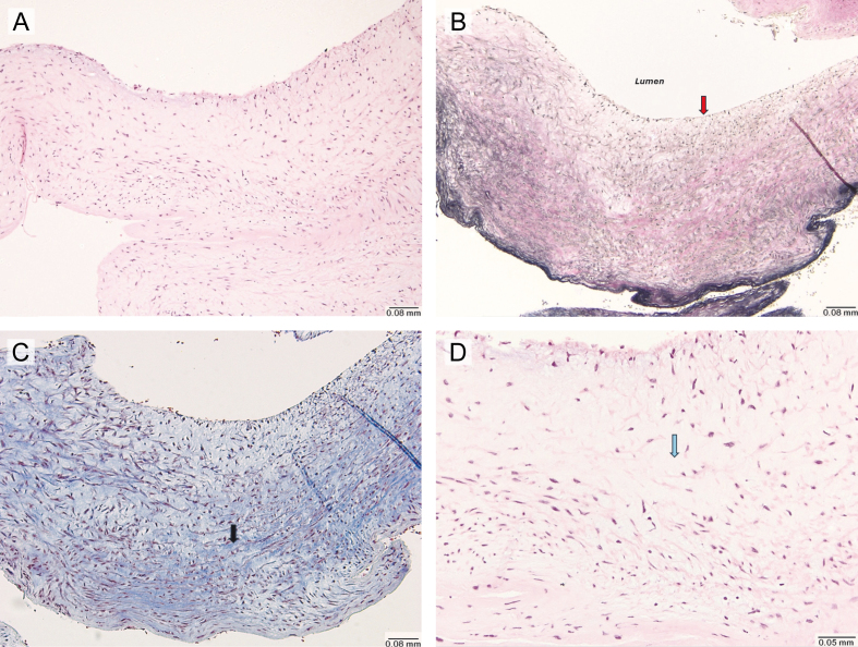
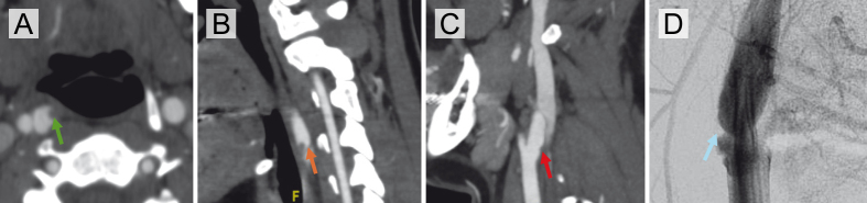
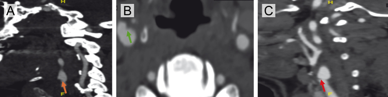
Background: Carotid artery webs (CWs) are an underrecognized cause of ischemic stroke, particularly in younger patients who lack conventional vascular risk factors. CWs are thought to represent an intimal variant of fibromuscular dysplasia (FMD); however, histopathologic data supporting this hypothesis remain limited. We report a case series of three patients with CW-related ischemic stroke who underwent carotid endarterectomy (CEA), allowing for histological analysis of the resected specimens. Methods: We retrospectively reviewed patients admitted to a Comprehensive Stroke Center between January 2015 and April 2025 with ischemic stroke or transient ischemic attack attributed to an ipsilateral carotid web who subsequently underwent carotid endarterectomy. Clinical data, imaging findings, and histopathologic features were analyzed. All cases met criteria for embolic stroke of undetermined source (ESUS) prior to surgery. Results: Three patients with CW-related stroke underwent carotid endarterectomy following recurrent events or high embolic risk. In two cases, superimposed thrombi led to initial misdiagnoses such as soft plaque or dissection. Histopathologic analysis consistently demonstrated fibrovascular tissue with intimal fibroid hyperplasia and myxoid degeneration, without lipid-rich plaques or inflammatory infiltrates. No patients experienced recurrent stroke or TIA by the time of their last documented follow-up. Conclusions: CWs represent a distinct non-atherosclerotic pathology characterized by intimal hyperplasia and myxoid degeneration. Superimposed thrombus may complicate diagnosis, often mimicking plaque or dissection. Advanced imaging, including MR vessel wall imaging and intravascular optical coherence tomography (OCT), can aid in accurate identification. Carotid revascularization may be effective in selected patients, particularly those with recurrence or ESUS. Prospective studies are needed to inform standardized diagnostic and therapeutic strategies.

 求助内容:
求助内容: 应助结果提醒方式:


