Edie Benedito Caetano, Luiz Angelo Vieira, Vinicius Santos Bueno, Túlio Stefanin Volpiani, Victor Hugo Monfrin Torres, Andrea Elisa Donovan Giraldo
{"title":"骨间前神经转移治疗桡神经损伤。","authors":"Edie Benedito Caetano, Luiz Angelo Vieira, Vinicius Santos Bueno, Túlio Stefanin Volpiani, Victor Hugo Monfrin Torres, Andrea Elisa Donovan Giraldo","doi":"10.1590/1413-785220253303e277311","DOIUrl":null,"url":null,"abstract":"<p><strong>Objectives: </strong>evaluate the anatomical characteristics and variations of the anterior interosseous nerve (AIN) and define the feasibility of transferring the pronator quadratus branch (PQB) to the posterior interosseous nerve (PIN) without tension.</p><p><strong>Materials and methods: </strong>Fifty upper limbs of 25 male adult cadavers were dissected, 20 were prepared and five were fresh cadavers.</p><p><strong>Results: </strong>The AIN originated from the median nerve in an average of 5.2 cm distal to the intercondylar line. In 29 limbs, it originated from the posterior fascicles of the median nerve, while in 21 specimens, from the posterolateral fascicles. In 2 specimens, two branches for the AIN were present. The PIN was studied in 30 limbs, we identified its origin in the radial nerve in all limbs. In 23 limbs, the branches to the supinator muscle originated from the PIN proximally to the Fröhse arcade, in 7 members distally.</p><p><strong>Conclusion: </strong>The PQB was reliably present in all dissected forearms and presented variations only in its diameter. The PQB could be transferred to PIN without tension in all specimens even with full range of motion of the forearm. <b><i>Level of Evidence IV; Case Series.</i></b></p>","PeriodicalId":55563,"journal":{"name":"Acta Ortopedica Brasileira","volume":"33 3","pages":"e277311"},"PeriodicalIF":0.6000,"publicationDate":"2025-08-18","publicationTypes":"Journal Article","fieldsOfStudy":null,"isOpenAccess":false,"openAccessPdf":"https://www.ncbi.nlm.nih.gov/pmc/articles/PMC12364506/pdf/","citationCount":"0","resultStr":"{\"title\":\"ANTERIOR INTEROSSEOUS NERVE TRANSFERS FOR THE TREATMENT OF RADIAL NERVE INJURIES.\",\"authors\":\"Edie Benedito Caetano, Luiz Angelo Vieira, Vinicius Santos Bueno, Túlio Stefanin Volpiani, Victor Hugo Monfrin Torres, Andrea Elisa Donovan Giraldo\",\"doi\":\"10.1590/1413-785220253303e277311\",\"DOIUrl\":null,\"url\":null,\"abstract\":\"<p><strong>Objectives: </strong>evaluate the anatomical characteristics and variations of the anterior interosseous nerve (AIN) and define the feasibility of transferring the pronator quadratus branch (PQB) to the posterior interosseous nerve (PIN) without tension.</p><p><strong>Materials and methods: </strong>Fifty upper limbs of 25 male adult cadavers were dissected, 20 were prepared and five were fresh cadavers.</p><p><strong>Results: </strong>The AIN originated from the median nerve in an average of 5.2 cm distal to the intercondylar line. In 29 limbs, it originated from the posterior fascicles of the median nerve, while in 21 specimens, from the posterolateral fascicles. In 2 specimens, two branches for the AIN were present. The PIN was studied in 30 limbs, we identified its origin in the radial nerve in all limbs. In 23 limbs, the branches to the supinator muscle originated from the PIN proximally to the Fröhse arcade, in 7 members distally.</p><p><strong>Conclusion: </strong>The PQB was reliably present in all dissected forearms and presented variations only in its diameter. The PQB could be transferred to PIN without tension in all specimens even with full range of motion of the forearm. <b><i>Level of Evidence IV; Case Series.</i></b></p>\",\"PeriodicalId\":55563,\"journal\":{\"name\":\"Acta Ortopedica Brasileira\",\"volume\":\"33 3\",\"pages\":\"e277311\"},\"PeriodicalIF\":0.6000,\"publicationDate\":\"2025-08-18\",\"publicationTypes\":\"Journal Article\",\"fieldsOfStudy\":null,\"isOpenAccess\":false,\"openAccessPdf\":\"https://www.ncbi.nlm.nih.gov/pmc/articles/PMC12364506/pdf/\",\"citationCount\":\"0\",\"resultStr\":null,\"platform\":\"Semanticscholar\",\"paperid\":null,\"PeriodicalName\":\"Acta Ortopedica Brasileira\",\"FirstCategoryId\":\"3\",\"ListUrlMain\":\"https://doi.org/10.1590/1413-785220253303e277311\",\"RegionNum\":4,\"RegionCategory\":\"医学\",\"ArticlePicture\":[],\"TitleCN\":null,\"AbstractTextCN\":null,\"PMCID\":null,\"EPubDate\":\"2025/1/1 0:00:00\",\"PubModel\":\"eCollection\",\"JCR\":\"Q4\",\"JCRName\":\"ORTHOPEDICS\",\"Score\":null,\"Total\":0}","platform":"Semanticscholar","paperid":null,"PeriodicalName":"Acta Ortopedica Brasileira","FirstCategoryId":"3","ListUrlMain":"https://doi.org/10.1590/1413-785220253303e277311","RegionNum":4,"RegionCategory":"医学","ArticlePicture":[],"TitleCN":null,"AbstractTextCN":null,"PMCID":null,"EPubDate":"2025/1/1 0:00:00","PubModel":"eCollection","JCR":"Q4","JCRName":"ORTHOPEDICS","Score":null,"Total":0}
ANTERIOR INTEROSSEOUS NERVE TRANSFERS FOR THE TREATMENT OF RADIAL NERVE INJURIES.
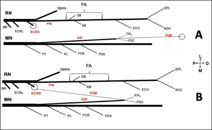
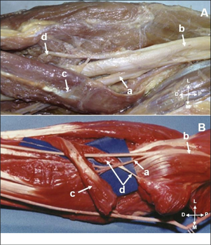
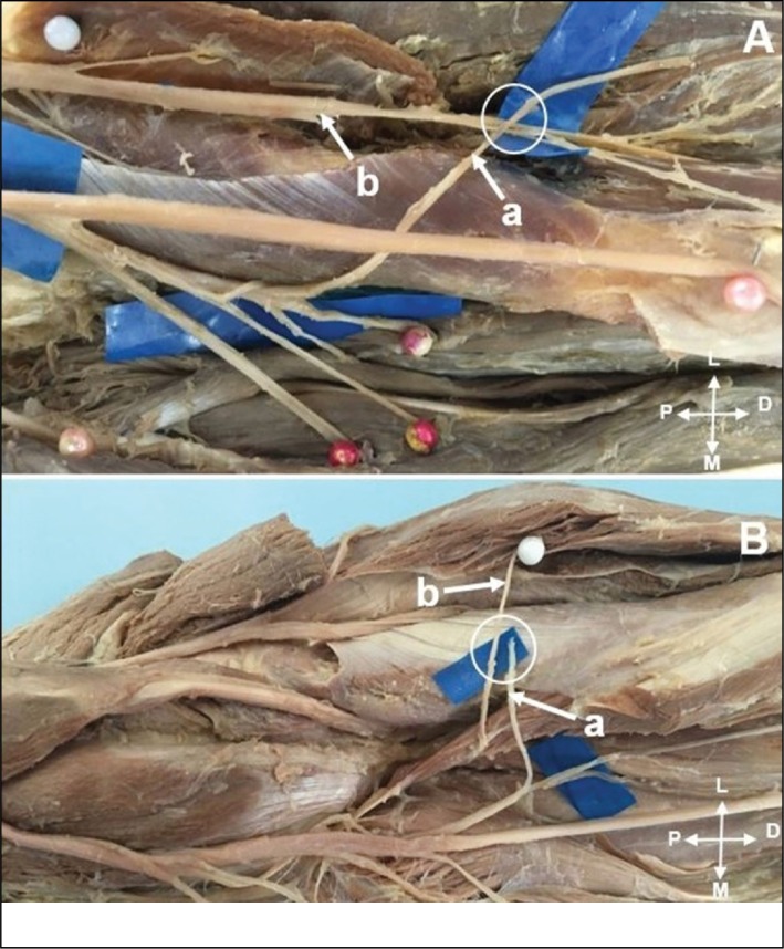
Objectives: evaluate the anatomical characteristics and variations of the anterior interosseous nerve (AIN) and define the feasibility of transferring the pronator quadratus branch (PQB) to the posterior interosseous nerve (PIN) without tension.
Materials and methods: Fifty upper limbs of 25 male adult cadavers were dissected, 20 were prepared and five were fresh cadavers.
Results: The AIN originated from the median nerve in an average of 5.2 cm distal to the intercondylar line. In 29 limbs, it originated from the posterior fascicles of the median nerve, while in 21 specimens, from the posterolateral fascicles. In 2 specimens, two branches for the AIN were present. The PIN was studied in 30 limbs, we identified its origin in the radial nerve in all limbs. In 23 limbs, the branches to the supinator muscle originated from the PIN proximally to the Fröhse arcade, in 7 members distally.
Conclusion: The PQB was reliably present in all dissected forearms and presented variations only in its diameter. The PQB could be transferred to PIN without tension in all specimens even with full range of motion of the forearm. Level of Evidence IV; Case Series.
期刊介绍:
A Revista Acta Ortopédica Brasileira, órgão oficial do Departamento de Ortopedia e Traumatologia da Faculdade de Medicina da Universidade de São Paulo (DOT/FMUSP), é publicada bimestralmente em seis edições ao ano (jan/fev, mar/abr, maio/jun, jul/ago, set/out e nov/dez) com versão em inglês disponível nos principais indexadores nacionais e internacionais e instituições de ensino do Brasil. Sendo hoje reconhecidamente uma importante contribuição para os especialistas da área com sua seriedade e árduo trabalho para as indexações já conquistadas.
 求助内容:
求助内容: 应助结果提醒方式:


