{"title":"多层螺旋ct引导下评价胶原-壳聚糖复合材料促进兔模型腕骨前关节融合术的作用。","authors":"Gannah-Samy, Alaa Samy, Awad Rizk, Emad Tolba, Zainab A Ramadan, Gamal Karrouf","doi":"10.1186/s13620-025-00307-1","DOIUrl":null,"url":null,"abstract":"<p><strong>Background: </strong>Arthrodesis is a critical procedure for restoring stability and relieving pain in severely damaged joints. Successful bone fusion remains a significant challenge, often necessitating the use of biomaterials to enhance healing. Collagen and chitosan, two natural polymers with established biocompatibility and osteoconductive properties, have shown promise in regenerative medicine applications. The present study aimed to evaluate the synergistic effect of a collagen-chitosan composite on bone fusion of the antebrachiocarpal joint in a rabbit model. Multislice CT morphometrical analysis was utilized to assess bone healing and fusion, offering detailed insights into the material's efficacy in promoting joint stabilization and bone regeneration.</p><p><strong>Materials and methods: </strong>Twelve healthy male New Zealand White rabbits (4.0 ± 0.3 months old) with a mean body weight of 2.5 ± 0.5 kg were used. These animals underwent curettage of the articular cartilage down to the subchondral bone. The rabbits were then randomly assigned into two groups: a control group (C), in which no composite was applied, and a treatment group, in which collagen-chitosan scaffolds were utilized (Col/Cs). Joint fusion was postoperatively assessed using a multislice detector computed tomography (MSCT).</p><p><strong>Result: </strong>(MSCT) revealed progressive enhancements in the collagen-chitosan (Col/Cs) group over 12 weeks. Radial cortical thickness and bone mineral density (BMD) were significantly higher at week 12 in the Col/Cs group (1.31 ± 0.10 mm vs. 1.03 ± 0.18 mm; p = 0.0086, and ~ 760 HU vs. ~510 HU; p = 0.0055, respectively). Intra-articular mineral density (IATMD) increased markedly at week 1 (p < 0.0001), decreased at week 6 (p < 0.0001), and rose again by week 12 (p < 0.0001), while the control group showed a gradual, non-significant increase. Joint space width decreased significantly in the Col/Cs group by week 6 (~ 0.6 mm vs. ~0.9 mm; p = 0.0034) and remained lower at week 12 (~ 0.55 mm vs. ~0.7 mm; p = 0.0062). Fusion ratio reached ~ 65% in the Col/Cs group compared to ~ 35% in controls (p < 0.0001). CBMD decreased in both groups by week 1 postoperatively but recovered more effectively in the Col/Cs group. By week 12, CBMD was significantly higher in the Col/Cs group (~ 1000 HU) than in controls (~ 950 HU; P < 0.0006). (UBMD) was initially similar (~ 780 HU), but by week 1, the Col/Cs group maintained higher values (~ 760 HU vs. ~620 HU; p < 0.0001), and this inclination continued through week 12 (~ 750 HU vs. ~680 HU; p = 0.001).</p><p><strong>Conclusion: </strong>The results of the present study indicate that the collagen-chitosan composite enhances bone fusion and joint stability in a rabbit model of antebrachiocarpal arthrodesis, demonstrating both innovation and potential clinical applicability.</p>","PeriodicalId":54916,"journal":{"name":"Irish Veterinary Journal","volume":"78 1","pages":"18"},"PeriodicalIF":3.1000,"publicationDate":"2025-08-26","publicationTypes":"Journal Article","fieldsOfStudy":null,"isOpenAccess":false,"openAccessPdf":"https://www.ncbi.nlm.nih.gov/pmc/articles/PMC12379432/pdf/","citationCount":"0","resultStr":"{\"title\":\"Multislice CT-guided evaluation of collagen-chitosan composite in promoting antebrachiocarpal arthrodesis in a rabbit model.\",\"authors\":\"Gannah-Samy, Alaa Samy, Awad Rizk, Emad Tolba, Zainab A Ramadan, Gamal Karrouf\",\"doi\":\"10.1186/s13620-025-00307-1\",\"DOIUrl\":null,\"url\":null,\"abstract\":\"<p><strong>Background: </strong>Arthrodesis is a critical procedure for restoring stability and relieving pain in severely damaged joints. Successful bone fusion remains a significant challenge, often necessitating the use of biomaterials to enhance healing. Collagen and chitosan, two natural polymers with established biocompatibility and osteoconductive properties, have shown promise in regenerative medicine applications. The present study aimed to evaluate the synergistic effect of a collagen-chitosan composite on bone fusion of the antebrachiocarpal joint in a rabbit model. Multislice CT morphometrical analysis was utilized to assess bone healing and fusion, offering detailed insights into the material's efficacy in promoting joint stabilization and bone regeneration.</p><p><strong>Materials and methods: </strong>Twelve healthy male New Zealand White rabbits (4.0 ± 0.3 months old) with a mean body weight of 2.5 ± 0.5 kg were used. These animals underwent curettage of the articular cartilage down to the subchondral bone. The rabbits were then randomly assigned into two groups: a control group (C), in which no composite was applied, and a treatment group, in which collagen-chitosan scaffolds were utilized (Col/Cs). Joint fusion was postoperatively assessed using a multislice detector computed tomography (MSCT).</p><p><strong>Result: </strong>(MSCT) revealed progressive enhancements in the collagen-chitosan (Col/Cs) group over 12 weeks. Radial cortical thickness and bone mineral density (BMD) were significantly higher at week 12 in the Col/Cs group (1.31 ± 0.10 mm vs. 1.03 ± 0.18 mm; p = 0.0086, and ~ 760 HU vs. ~510 HU; p = 0.0055, respectively). Intra-articular mineral density (IATMD) increased markedly at week 1 (p < 0.0001), decreased at week 6 (p < 0.0001), and rose again by week 12 (p < 0.0001), while the control group showed a gradual, non-significant increase. Joint space width decreased significantly in the Col/Cs group by week 6 (~ 0.6 mm vs. ~0.9 mm; p = 0.0034) and remained lower at week 12 (~ 0.55 mm vs. ~0.7 mm; p = 0.0062). Fusion ratio reached ~ 65% in the Col/Cs group compared to ~ 35% in controls (p < 0.0001). CBMD decreased in both groups by week 1 postoperatively but recovered more effectively in the Col/Cs group. By week 12, CBMD was significantly higher in the Col/Cs group (~ 1000 HU) than in controls (~ 950 HU; P < 0.0006). (UBMD) was initially similar (~ 780 HU), but by week 1, the Col/Cs group maintained higher values (~ 760 HU vs. ~620 HU; p < 0.0001), and this inclination continued through week 12 (~ 750 HU vs. ~680 HU; p = 0.001).</p><p><strong>Conclusion: </strong>The results of the present study indicate that the collagen-chitosan composite enhances bone fusion and joint stability in a rabbit model of antebrachiocarpal arthrodesis, demonstrating both innovation and potential clinical applicability.</p>\",\"PeriodicalId\":54916,\"journal\":{\"name\":\"Irish Veterinary Journal\",\"volume\":\"78 1\",\"pages\":\"18\"},\"PeriodicalIF\":3.1000,\"publicationDate\":\"2025-08-26\",\"publicationTypes\":\"Journal Article\",\"fieldsOfStudy\":null,\"isOpenAccess\":false,\"openAccessPdf\":\"https://www.ncbi.nlm.nih.gov/pmc/articles/PMC12379432/pdf/\",\"citationCount\":\"0\",\"resultStr\":null,\"platform\":\"Semanticscholar\",\"paperid\":null,\"PeriodicalName\":\"Irish Veterinary Journal\",\"FirstCategoryId\":\"97\",\"ListUrlMain\":\"https://doi.org/10.1186/s13620-025-00307-1\",\"RegionNum\":2,\"RegionCategory\":\"农林科学\",\"ArticlePicture\":[],\"TitleCN\":null,\"AbstractTextCN\":null,\"PMCID\":null,\"EPubDate\":\"\",\"PubModel\":\"\",\"JCR\":\"Q1\",\"JCRName\":\"VETERINARY SCIENCES\",\"Score\":null,\"Total\":0}","platform":"Semanticscholar","paperid":null,"PeriodicalName":"Irish Veterinary Journal","FirstCategoryId":"97","ListUrlMain":"https://doi.org/10.1186/s13620-025-00307-1","RegionNum":2,"RegionCategory":"农林科学","ArticlePicture":[],"TitleCN":null,"AbstractTextCN":null,"PMCID":null,"EPubDate":"","PubModel":"","JCR":"Q1","JCRName":"VETERINARY SCIENCES","Score":null,"Total":0}
引用次数: 0
摘要
背景:关节融合术是恢复严重损伤关节稳定性和缓解疼痛的关键手术。成功的骨融合仍然是一个重大挑战,通常需要使用生物材料来增强愈合。胶原蛋白和壳聚糖是两种具有生物相容性和骨导电性的天然聚合物,在再生医学中有着广阔的应用前景。本研究旨在评价胶原-壳聚糖复合材料对兔腕前关节骨融合的协同作用。多层螺旋CT形态测量分析用于评估骨愈合和融合,为材料促进关节稳定和骨再生的功效提供详细的见解。材料与方法:选用健康雄性新西兰大白兔12只(4.0±0.3月龄),平均体重2.5±0.5 kg。这些动物的关节软骨被刮除至软骨下骨。然后将家兔随机分为两组:对照组(C)不使用复合材料,治疗组(Col/Cs)使用胶原-壳聚糖支架。术后采用多层计算机断层扫描(MSCT)评估关节融合情况。结果:MSCT显示胶原-壳聚糖(Col/Cs)组在12周内呈进行性增强。Col/Cs组桡骨皮质厚度和骨密度(BMD)在第12周显著升高(1.31±0.10 mm vs 1.03±0.18 mm, p = 0.0086, ~ 760 HU vs ~510 HU, p = 0.0055)。结论:本研究结果表明,胶原-壳聚糖复合材料可增强兔腕前关节融合术模型的骨融合和关节稳定性,具有创新性和潜在的临床应用价值。
Multislice CT-guided evaluation of collagen-chitosan composite in promoting antebrachiocarpal arthrodesis in a rabbit model.
Background: Arthrodesis is a critical procedure for restoring stability and relieving pain in severely damaged joints. Successful bone fusion remains a significant challenge, often necessitating the use of biomaterials to enhance healing. Collagen and chitosan, two natural polymers with established biocompatibility and osteoconductive properties, have shown promise in regenerative medicine applications. The present study aimed to evaluate the synergistic effect of a collagen-chitosan composite on bone fusion of the antebrachiocarpal joint in a rabbit model. Multislice CT morphometrical analysis was utilized to assess bone healing and fusion, offering detailed insights into the material's efficacy in promoting joint stabilization and bone regeneration.
Materials and methods: Twelve healthy male New Zealand White rabbits (4.0 ± 0.3 months old) with a mean body weight of 2.5 ± 0.5 kg were used. These animals underwent curettage of the articular cartilage down to the subchondral bone. The rabbits were then randomly assigned into two groups: a control group (C), in which no composite was applied, and a treatment group, in which collagen-chitosan scaffolds were utilized (Col/Cs). Joint fusion was postoperatively assessed using a multislice detector computed tomography (MSCT).
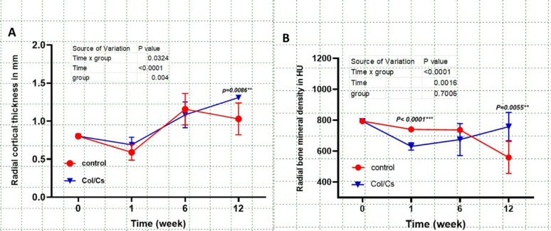
Result: (MSCT) revealed progressive enhancements in the collagen-chitosan (Col/Cs) group over 12 weeks. Radial cortical thickness and bone mineral density (BMD) were significantly higher at week 12 in the Col/Cs group (1.31 ± 0.10 mm vs. 1.03 ± 0.18 mm; p = 0.0086, and ~ 760 HU vs. ~510 HU; p = 0.0055, respectively). Intra-articular mineral density (IATMD) increased markedly at week 1 (p < 0.0001), decreased at week 6 (p < 0.0001), and rose again by week 12 (p < 0.0001), while the control group showed a gradual, non-significant increase. Joint space width decreased significantly in the Col/Cs group by week 6 (~ 0.6 mm vs. ~0.9 mm; p = 0.0034) and remained lower at week 12 (~ 0.55 mm vs. ~0.7 mm; p = 0.0062). Fusion ratio reached ~ 65% in the Col/Cs group compared to ~ 35% in controls (p < 0.0001). CBMD decreased in both groups by week 1 postoperatively but recovered more effectively in the Col/Cs group. By week 12, CBMD was significantly higher in the Col/Cs group (~ 1000 HU) than in controls (~ 950 HU; P < 0.0006). (UBMD) was initially similar (~ 780 HU), but by week 1, the Col/Cs group maintained higher values (~ 760 HU vs. ~620 HU; p < 0.0001), and this inclination continued through week 12 (~ 750 HU vs. ~680 HU; p = 0.001).
Conclusion: The results of the present study indicate that the collagen-chitosan composite enhances bone fusion and joint stability in a rabbit model of antebrachiocarpal arthrodesis, demonstrating both innovation and potential clinical applicability.
期刊介绍:
Irish Veterinary Journal is an open access journal with a vision to make a substantial contribution to the dissemination of evidence-based knowledge that will promote optimal health and welfare of both domestic and wild species of animals.
Irish Veterinary Journal has a clinical research focus with an emphasis on the effective management of health in both individual and populations of animals. Published studies will be relevant to both the international veterinary profession and veterinary scientists. Papers relating to veterinary education, veterinary ethics, veterinary public health, or relevant studies in the area of social science (participatory research) are also within the scope of Irish Veterinary Journal.
 求助内容:
求助内容: 应助结果提醒方式:


