Weixia Li, Yajing Zhu, Gangde Zhao, Xiaoyan Chen, Xiangtian Zhao, Haimin Xu, Yingyu Che, Yinan Chen, Yuxiang Ye, Xin Dou, Hui Wang, Jingliang Cheng, Qing Xie, Kemin Chen
{"title":"基于深度学习的基于磁共振图像和非图像数据的肝纤维化自动评估。","authors":"Weixia Li, Yajing Zhu, Gangde Zhao, Xiaoyan Chen, Xiangtian Zhao, Haimin Xu, Yingyu Che, Yinan Chen, Yuxiang Ye, Xin Dou, Hui Wang, Jingliang Cheng, Qing Xie, Kemin Chen","doi":"10.21037/qims-2024-2506","DOIUrl":null,"url":null,"abstract":"<p><strong>Background: </strong>Accurate staging of hepatic fibrosis is critical for prognostication and management among patients with chronic liver disease, and noninvasive, efficient alternatives to biopsy are urgently needed. This study aimed to evaluate the performance of an automated deep learning (DL) algorithm for fibrosis staging and for differentiating patients with hepatic fibrosis from healthy individuals via magnetic resonance (MR) images with and without additional clinical data.</p><p><strong>Methods: </strong>A total of 500 patients from two medical centers were retrospectively analyzed. DL models were developed based on delayed-phase MR images to predict fibrosis stages. Additional models were constructed by integrating the DL algorithm with nonimaging variables, including serologic biomarkers [aminotransferase-to-platelet ratio index (APRI) and fibrosis index based on four factors (FIB-4)], viral status (hepatitis B and C), and MR scanner parameters. Diagnostic performance, was assessed via the area under the receiver operating characteristic curve (AUROC), and comparisons were through use of the DeLong test. Sensitivity and specificity of the DL and full models (DL plus all clinical features) were compared with those of experienced radiologists and serologic biomarkers via the McNemar test.</p><p><strong>Results: </strong>In the test set, the full model achieved AUROC values of 0.99 [95% confidence interval (CI): 0.94-1.00], 0.98 (95% CI: 0.93-0.99), 0.90 (95% CI: 0.83-0.95), 0.81 (95% CI: 0.73-0.88), and 0.84 (95% CI: 0.76-0.90) for staging F0-4, F1-4, F2-4, F3-4, and F4, respectively. This model significantly outperformed the DL model in early-stage classification (F0-4 and F1-4). Compared with expert radiologists, it showed superior specificity for F0-4 and higher sensitivity across the other four classification tasks. Both the DL and full models showed significantly greater specificity than did the biomarkers for staging advanced fibrosis (F3-4 and F4).</p><p><strong>Conclusions: </strong>The proposed DL algorithm provides a noninvasive method for hepatic fibrosis staging and screening, outperforming both radiologists and conventional biomarkers, and may facilitate improved clinical decision-making.</p>","PeriodicalId":54267,"journal":{"name":"Quantitative Imaging in Medicine and Surgery","volume":"15 9","pages":"8250-8264"},"PeriodicalIF":2.3000,"publicationDate":"2025-09-01","publicationTypes":"Journal Article","fieldsOfStudy":null,"isOpenAccess":false,"openAccessPdf":"https://www.ncbi.nlm.nih.gov/pmc/articles/PMC12397629/pdf/","citationCount":"0","resultStr":"{\"title\":\"Deep learning-based automated assessment of hepatic fibrosis via magnetic resonance images and nonimage data.\",\"authors\":\"Weixia Li, Yajing Zhu, Gangde Zhao, Xiaoyan Chen, Xiangtian Zhao, Haimin Xu, Yingyu Che, Yinan Chen, Yuxiang Ye, Xin Dou, Hui Wang, Jingliang Cheng, Qing Xie, Kemin Chen\",\"doi\":\"10.21037/qims-2024-2506\",\"DOIUrl\":null,\"url\":null,\"abstract\":\"<p><strong>Background: </strong>Accurate staging of hepatic fibrosis is critical for prognostication and management among patients with chronic liver disease, and noninvasive, efficient alternatives to biopsy are urgently needed. This study aimed to evaluate the performance of an automated deep learning (DL) algorithm for fibrosis staging and for differentiating patients with hepatic fibrosis from healthy individuals via magnetic resonance (MR) images with and without additional clinical data.</p><p><strong>Methods: </strong>A total of 500 patients from two medical centers were retrospectively analyzed. DL models were developed based on delayed-phase MR images to predict fibrosis stages. Additional models were constructed by integrating the DL algorithm with nonimaging variables, including serologic biomarkers [aminotransferase-to-platelet ratio index (APRI) and fibrosis index based on four factors (FIB-4)], viral status (hepatitis B and C), and MR scanner parameters. Diagnostic performance, was assessed via the area under the receiver operating characteristic curve (AUROC), and comparisons were through use of the DeLong test. Sensitivity and specificity of the DL and full models (DL plus all clinical features) were compared with those of experienced radiologists and serologic biomarkers via the McNemar test.</p><p><strong>Results: </strong>In the test set, the full model achieved AUROC values of 0.99 [95% confidence interval (CI): 0.94-1.00], 0.98 (95% CI: 0.93-0.99), 0.90 (95% CI: 0.83-0.95), 0.81 (95% CI: 0.73-0.88), and 0.84 (95% CI: 0.76-0.90) for staging F0-4, F1-4, F2-4, F3-4, and F4, respectively. This model significantly outperformed the DL model in early-stage classification (F0-4 and F1-4). Compared with expert radiologists, it showed superior specificity for F0-4 and higher sensitivity across the other four classification tasks. Both the DL and full models showed significantly greater specificity than did the biomarkers for staging advanced fibrosis (F3-4 and F4).</p><p><strong>Conclusions: </strong>The proposed DL algorithm provides a noninvasive method for hepatic fibrosis staging and screening, outperforming both radiologists and conventional biomarkers, and may facilitate improved clinical decision-making.</p>\",\"PeriodicalId\":54267,\"journal\":{\"name\":\"Quantitative Imaging in Medicine and Surgery\",\"volume\":\"15 9\",\"pages\":\"8250-8264\"},\"PeriodicalIF\":2.3000,\"publicationDate\":\"2025-09-01\",\"publicationTypes\":\"Journal Article\",\"fieldsOfStudy\":null,\"isOpenAccess\":false,\"openAccessPdf\":\"https://www.ncbi.nlm.nih.gov/pmc/articles/PMC12397629/pdf/\",\"citationCount\":\"0\",\"resultStr\":null,\"platform\":\"Semanticscholar\",\"paperid\":null,\"PeriodicalName\":\"Quantitative Imaging in Medicine and Surgery\",\"FirstCategoryId\":\"3\",\"ListUrlMain\":\"https://doi.org/10.21037/qims-2024-2506\",\"RegionNum\":2,\"RegionCategory\":\"医学\",\"ArticlePicture\":[],\"TitleCN\":null,\"AbstractTextCN\":null,\"PMCID\":null,\"EPubDate\":\"2025/8/18 0:00:00\",\"PubModel\":\"Epub\",\"JCR\":\"Q2\",\"JCRName\":\"RADIOLOGY, NUCLEAR MEDICINE & MEDICAL IMAGING\",\"Score\":null,\"Total\":0}","platform":"Semanticscholar","paperid":null,"PeriodicalName":"Quantitative Imaging in Medicine and Surgery","FirstCategoryId":"3","ListUrlMain":"https://doi.org/10.21037/qims-2024-2506","RegionNum":2,"RegionCategory":"医学","ArticlePicture":[],"TitleCN":null,"AbstractTextCN":null,"PMCID":null,"EPubDate":"2025/8/18 0:00:00","PubModel":"Epub","JCR":"Q2","JCRName":"RADIOLOGY, NUCLEAR MEDICINE & MEDICAL IMAGING","Score":null,"Total":0}
Deep learning-based automated assessment of hepatic fibrosis via magnetic resonance images and nonimage data.
Background: Accurate staging of hepatic fibrosis is critical for prognostication and management among patients with chronic liver disease, and noninvasive, efficient alternatives to biopsy are urgently needed. This study aimed to evaluate the performance of an automated deep learning (DL) algorithm for fibrosis staging and for differentiating patients with hepatic fibrosis from healthy individuals via magnetic resonance (MR) images with and without additional clinical data.
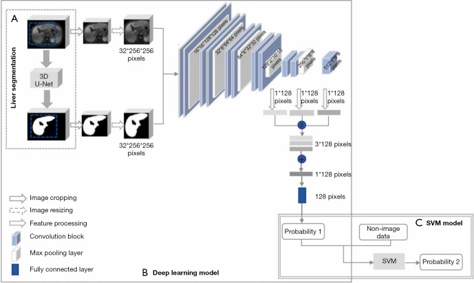
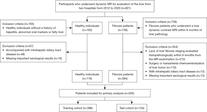
Methods: A total of 500 patients from two medical centers were retrospectively analyzed. DL models were developed based on delayed-phase MR images to predict fibrosis stages. Additional models were constructed by integrating the DL algorithm with nonimaging variables, including serologic biomarkers [aminotransferase-to-platelet ratio index (APRI) and fibrosis index based on four factors (FIB-4)], viral status (hepatitis B and C), and MR scanner parameters. Diagnostic performance, was assessed via the area under the receiver operating characteristic curve (AUROC), and comparisons were through use of the DeLong test. Sensitivity and specificity of the DL and full models (DL plus all clinical features) were compared with those of experienced radiologists and serologic biomarkers via the McNemar test.
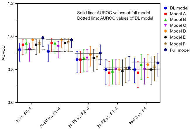
Results: In the test set, the full model achieved AUROC values of 0.99 [95% confidence interval (CI): 0.94-1.00], 0.98 (95% CI: 0.93-0.99), 0.90 (95% CI: 0.83-0.95), 0.81 (95% CI: 0.73-0.88), and 0.84 (95% CI: 0.76-0.90) for staging F0-4, F1-4, F2-4, F3-4, and F4, respectively. This model significantly outperformed the DL model in early-stage classification (F0-4 and F1-4). Compared with expert radiologists, it showed superior specificity for F0-4 and higher sensitivity across the other four classification tasks. Both the DL and full models showed significantly greater specificity than did the biomarkers for staging advanced fibrosis (F3-4 and F4).
Conclusions: The proposed DL algorithm provides a noninvasive method for hepatic fibrosis staging and screening, outperforming both radiologists and conventional biomarkers, and may facilitate improved clinical decision-making.

 求助内容:
求助内容: 应助结果提醒方式:


