{"title":"术前多序列磁共振成像栖息地分析预测脑膜瘤分级。","authors":"Zongyou Cai, Ye Heng Wong, Tiffany Y So","doi":"10.21037/qims-2025-1041","DOIUrl":null,"url":null,"abstract":"<p><strong>Background: </strong>Accurate grading of meningiomas is crucial for patient prognostication and management. Intratumoral heterogeneity may lead to differences in the biological and radiological properties observed within different tumor subregions. This study aimed to represent the spatial distributions and local patterns of tumor heterogeneity in meningiomas using non-invasive habitat analysis on filtered multisequence magnetic resonance imaging (MRI) and evaluate the utility of integrated models combining habitat and clinical data for meningioma grade prediction.</p><p><strong>Methods: </strong>Sixty patients with pathologically confirmed meningiomas [30 World Health Organization (WHO) grade 1, 28 grade 2, 2 grade 3] were retrospectively included in this cross-sectional study. Pre-operative T2-weighted (T2W) and T1-weighted with contrast (T1C) MRI sequences were processed using a three-dimensional (3D) Laplacian of Gaussian (LoG) filter (σ=3), and four distinct tumor habitats were generated using Otsu's thresholding method. Relative mean, relative standard deviation (SD), and entropy were quantified for each habitat on MRI.</p><p><strong>Results: </strong>Significant differences in relative mean intensities were observed between habitats in individual patients for both low-grade and high-grade meningiomas (P<0.01). High-grade meningiomas exhibited significantly higher relative mean and SD of T2W and T1C intensities across habitats compared to low-grade tumors (P≤0.03). The entropy of T1C was also significantly higher in high-grade tumors (P≤0.01). The integrated model incorporating the selected habitat measures and clinical factors achieved an area under the curve (AUC) of 0.84 [95% bootstrap confidence interval (CI): 0.72-0.92] in differentiating high-grade from low-grade meningiomas, with 0.78 accuracy, 0.73 sensitivity, and 0.83 specificity.</p><p><strong>Conclusions: </strong>Habitat analysis of conventional multisequence MRI provides a promising non-invasive approach to capture tumor heterogeneity for meningioma grading.</p>","PeriodicalId":54267,"journal":{"name":"Quantitative Imaging in Medicine and Surgery","volume":"15 9","pages":"7874-7884"},"PeriodicalIF":2.3000,"publicationDate":"2025-09-01","publicationTypes":"Journal Article","fieldsOfStudy":null,"isOpenAccess":false,"openAccessPdf":"https://www.ncbi.nlm.nih.gov/pmc/articles/PMC12397627/pdf/","citationCount":"0","resultStr":"{\"title\":\"Multisequence magnetic resonance imaging habitat analysis for pre-operative meningioma grade prediction.\",\"authors\":\"Zongyou Cai, Ye Heng Wong, Tiffany Y So\",\"doi\":\"10.21037/qims-2025-1041\",\"DOIUrl\":null,\"url\":null,\"abstract\":\"<p><strong>Background: </strong>Accurate grading of meningiomas is crucial for patient prognostication and management. Intratumoral heterogeneity may lead to differences in the biological and radiological properties observed within different tumor subregions. This study aimed to represent the spatial distributions and local patterns of tumor heterogeneity in meningiomas using non-invasive habitat analysis on filtered multisequence magnetic resonance imaging (MRI) and evaluate the utility of integrated models combining habitat and clinical data for meningioma grade prediction.</p><p><strong>Methods: </strong>Sixty patients with pathologically confirmed meningiomas [30 World Health Organization (WHO) grade 1, 28 grade 2, 2 grade 3] were retrospectively included in this cross-sectional study. Pre-operative T2-weighted (T2W) and T1-weighted with contrast (T1C) MRI sequences were processed using a three-dimensional (3D) Laplacian of Gaussian (LoG) filter (σ=3), and four distinct tumor habitats were generated using Otsu's thresholding method. Relative mean, relative standard deviation (SD), and entropy were quantified for each habitat on MRI.</p><p><strong>Results: </strong>Significant differences in relative mean intensities were observed between habitats in individual patients for both low-grade and high-grade meningiomas (P<0.01). High-grade meningiomas exhibited significantly higher relative mean and SD of T2W and T1C intensities across habitats compared to low-grade tumors (P≤0.03). The entropy of T1C was also significantly higher in high-grade tumors (P≤0.01). The integrated model incorporating the selected habitat measures and clinical factors achieved an area under the curve (AUC) of 0.84 [95% bootstrap confidence interval (CI): 0.72-0.92] in differentiating high-grade from low-grade meningiomas, with 0.78 accuracy, 0.73 sensitivity, and 0.83 specificity.</p><p><strong>Conclusions: </strong>Habitat analysis of conventional multisequence MRI provides a promising non-invasive approach to capture tumor heterogeneity for meningioma grading.</p>\",\"PeriodicalId\":54267,\"journal\":{\"name\":\"Quantitative Imaging in Medicine and Surgery\",\"volume\":\"15 9\",\"pages\":\"7874-7884\"},\"PeriodicalIF\":2.3000,\"publicationDate\":\"2025-09-01\",\"publicationTypes\":\"Journal Article\",\"fieldsOfStudy\":null,\"isOpenAccess\":false,\"openAccessPdf\":\"https://www.ncbi.nlm.nih.gov/pmc/articles/PMC12397627/pdf/\",\"citationCount\":\"0\",\"resultStr\":null,\"platform\":\"Semanticscholar\",\"paperid\":null,\"PeriodicalName\":\"Quantitative Imaging in Medicine and Surgery\",\"FirstCategoryId\":\"3\",\"ListUrlMain\":\"https://doi.org/10.21037/qims-2025-1041\",\"RegionNum\":2,\"RegionCategory\":\"医学\",\"ArticlePicture\":[],\"TitleCN\":null,\"AbstractTextCN\":null,\"PMCID\":null,\"EPubDate\":\"2025/8/19 0:00:00\",\"PubModel\":\"Epub\",\"JCR\":\"Q2\",\"JCRName\":\"RADIOLOGY, NUCLEAR MEDICINE & MEDICAL IMAGING\",\"Score\":null,\"Total\":0}","platform":"Semanticscholar","paperid":null,"PeriodicalName":"Quantitative Imaging in Medicine and Surgery","FirstCategoryId":"3","ListUrlMain":"https://doi.org/10.21037/qims-2025-1041","RegionNum":2,"RegionCategory":"医学","ArticlePicture":[],"TitleCN":null,"AbstractTextCN":null,"PMCID":null,"EPubDate":"2025/8/19 0:00:00","PubModel":"Epub","JCR":"Q2","JCRName":"RADIOLOGY, NUCLEAR MEDICINE & MEDICAL IMAGING","Score":null,"Total":0}
Multisequence magnetic resonance imaging habitat analysis for pre-operative meningioma grade prediction.
Background: Accurate grading of meningiomas is crucial for patient prognostication and management. Intratumoral heterogeneity may lead to differences in the biological and radiological properties observed within different tumor subregions. This study aimed to represent the spatial distributions and local patterns of tumor heterogeneity in meningiomas using non-invasive habitat analysis on filtered multisequence magnetic resonance imaging (MRI) and evaluate the utility of integrated models combining habitat and clinical data for meningioma grade prediction.
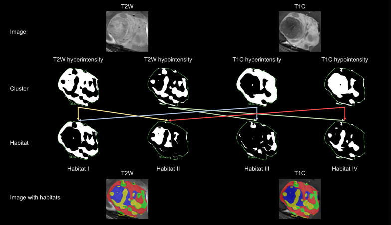
Methods: Sixty patients with pathologically confirmed meningiomas [30 World Health Organization (WHO) grade 1, 28 grade 2, 2 grade 3] were retrospectively included in this cross-sectional study. Pre-operative T2-weighted (T2W) and T1-weighted with contrast (T1C) MRI sequences were processed using a three-dimensional (3D) Laplacian of Gaussian (LoG) filter (σ=3), and four distinct tumor habitats were generated using Otsu's thresholding method. Relative mean, relative standard deviation (SD), and entropy were quantified for each habitat on MRI.
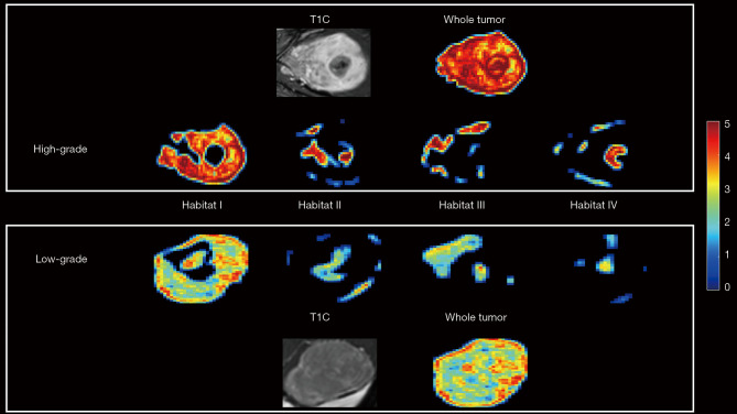
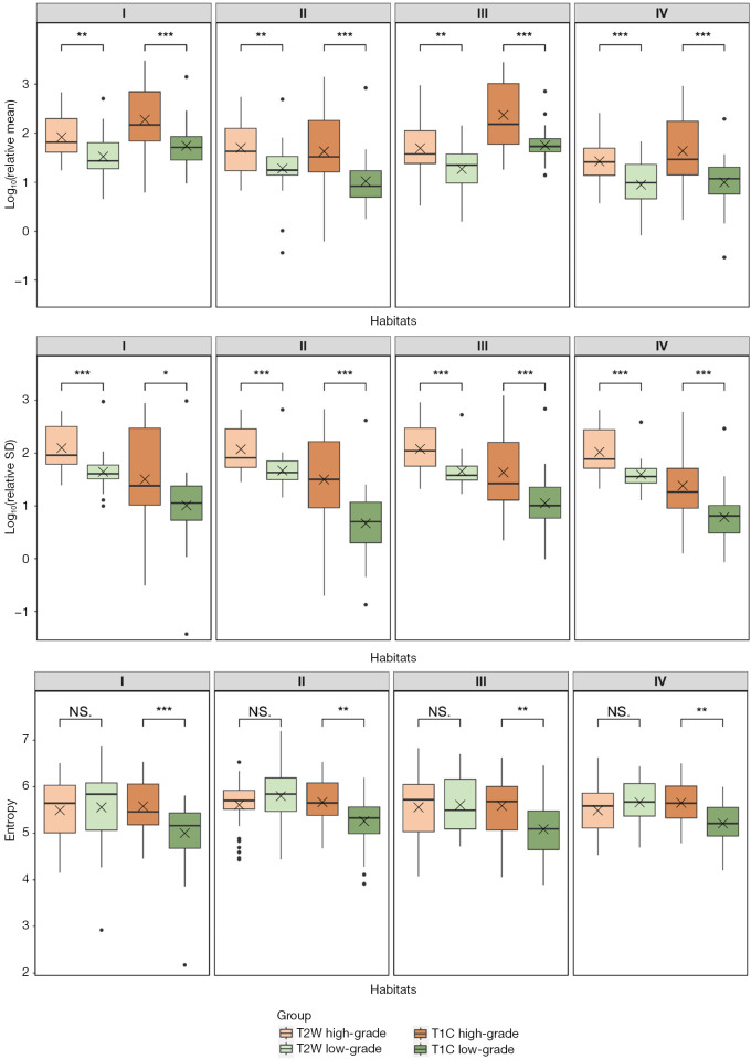
Results: Significant differences in relative mean intensities were observed between habitats in individual patients for both low-grade and high-grade meningiomas (P<0.01). High-grade meningiomas exhibited significantly higher relative mean and SD of T2W and T1C intensities across habitats compared to low-grade tumors (P≤0.03). The entropy of T1C was also significantly higher in high-grade tumors (P≤0.01). The integrated model incorporating the selected habitat measures and clinical factors achieved an area under the curve (AUC) of 0.84 [95% bootstrap confidence interval (CI): 0.72-0.92] in differentiating high-grade from low-grade meningiomas, with 0.78 accuracy, 0.73 sensitivity, and 0.83 specificity.
Conclusions: Habitat analysis of conventional multisequence MRI provides a promising non-invasive approach to capture tumor heterogeneity for meningioma grading.

 求助内容:
求助内容: 应助结果提醒方式:


