{"title":"光学相干断层摄影血管造影定量分析免疫球蛋白A肾病患者黄斑和视盘微血管参数。","authors":"Chen Qiu, Guihong Wu, Xueqin Zhang, Ting Kang, Dongmei Zhang, Mengxia Cao, Shuhan Zeng, Rui Wang, Tianyu Yao, Jie Chen, Santao Ou, Yue He","doi":"10.21037/qims-24-2113","DOIUrl":null,"url":null,"abstract":"<p><strong>Background: </strong>Immunoglobulin A nephropathy (IgAN) is a common chronic glomerulonephritis. The kidneys and eyes have structural and developmental similarities, but ocular microvascular changes in IgAN, especially in the macular and optic disc, are poorly studied. This study aimed to assess these microvascular parameters in IgAN patients using optical coherence tomography (OCT) and OCT angiography (OCTA) and explore their associations with clinical data.</p><p><strong>Methods: </strong>This is a case-control study. Fifteen patients with IgAN and 15 healthy controls (HCs) who attended The Affiliated Hospital of Southwest Medical University between July 2022 and December 2023 were selected. OCT and OCTA were used to assess macular thickness, retinal nerve fiber layer (RNFL) thickness, the vascular density (VD) and perfusion density (PD) of macular and optic disc, and foveal avascular zone (FAZ). In addition, we analyzed the association between OCT and OCTA parameters and clinical data in patients with IgAN.</p><p><strong>Results: </strong>VD and PD were significantly lower in the macular area in the IgAN group than the HCs (VD: 18.270±0.683 <i>vs.</i> 18.943±0.357, P<0.001; PD: 0.447±0.019 <i>vs.</i> 0.464±0.009, P<0.001). VD and PD in the macular area were negatively correlated with urine protein levels (VD: ρ=-0.604, P=0.017; PD: ρ=-0.551, P=0.033) and urine microalbumin levels (VD: ρ=-0.762, P=0.001; PD: ρ=-0.751, P=0.001). The FAZ area was positively correlated with 24-h urine volume (ρ=0.651, P=0.009).</p><p><strong>Conclusions: </strong>Compared with HCs, the status of macular microcirculation in the IgAN group was significantly decreased, and some microvascular parameters were significantly correlated with the clinical data.</p>","PeriodicalId":54267,"journal":{"name":"Quantitative Imaging in Medicine and Surgery","volume":"15 9","pages":"7803-7819"},"PeriodicalIF":2.3000,"publicationDate":"2025-09-01","publicationTypes":"Journal Article","fieldsOfStudy":null,"isOpenAccess":false,"openAccessPdf":"https://www.ncbi.nlm.nih.gov/pmc/articles/PMC12397639/pdf/","citationCount":"0","resultStr":"{\"title\":\"Quantitative analysis of macular and optic disc microvascular parameters in patients with immunoglobulin A nephropathy using optical coherence tomography angiography.\",\"authors\":\"Chen Qiu, Guihong Wu, Xueqin Zhang, Ting Kang, Dongmei Zhang, Mengxia Cao, Shuhan Zeng, Rui Wang, Tianyu Yao, Jie Chen, Santao Ou, Yue He\",\"doi\":\"10.21037/qims-24-2113\",\"DOIUrl\":null,\"url\":null,\"abstract\":\"<p><strong>Background: </strong>Immunoglobulin A nephropathy (IgAN) is a common chronic glomerulonephritis. The kidneys and eyes have structural and developmental similarities, but ocular microvascular changes in IgAN, especially in the macular and optic disc, are poorly studied. This study aimed to assess these microvascular parameters in IgAN patients using optical coherence tomography (OCT) and OCT angiography (OCTA) and explore their associations with clinical data.</p><p><strong>Methods: </strong>This is a case-control study. Fifteen patients with IgAN and 15 healthy controls (HCs) who attended The Affiliated Hospital of Southwest Medical University between July 2022 and December 2023 were selected. OCT and OCTA were used to assess macular thickness, retinal nerve fiber layer (RNFL) thickness, the vascular density (VD) and perfusion density (PD) of macular and optic disc, and foveal avascular zone (FAZ). In addition, we analyzed the association between OCT and OCTA parameters and clinical data in patients with IgAN.</p><p><strong>Results: </strong>VD and PD were significantly lower in the macular area in the IgAN group than the HCs (VD: 18.270±0.683 <i>vs.</i> 18.943±0.357, P<0.001; PD: 0.447±0.019 <i>vs.</i> 0.464±0.009, P<0.001). VD and PD in the macular area were negatively correlated with urine protein levels (VD: ρ=-0.604, P=0.017; PD: ρ=-0.551, P=0.033) and urine microalbumin levels (VD: ρ=-0.762, P=0.001; PD: ρ=-0.751, P=0.001). The FAZ area was positively correlated with 24-h urine volume (ρ=0.651, P=0.009).</p><p><strong>Conclusions: </strong>Compared with HCs, the status of macular microcirculation in the IgAN group was significantly decreased, and some microvascular parameters were significantly correlated with the clinical data.</p>\",\"PeriodicalId\":54267,\"journal\":{\"name\":\"Quantitative Imaging in Medicine and Surgery\",\"volume\":\"15 9\",\"pages\":\"7803-7819\"},\"PeriodicalIF\":2.3000,\"publicationDate\":\"2025-09-01\",\"publicationTypes\":\"Journal Article\",\"fieldsOfStudy\":null,\"isOpenAccess\":false,\"openAccessPdf\":\"https://www.ncbi.nlm.nih.gov/pmc/articles/PMC12397639/pdf/\",\"citationCount\":\"0\",\"resultStr\":null,\"platform\":\"Semanticscholar\",\"paperid\":null,\"PeriodicalName\":\"Quantitative Imaging in Medicine and Surgery\",\"FirstCategoryId\":\"3\",\"ListUrlMain\":\"https://doi.org/10.21037/qims-24-2113\",\"RegionNum\":2,\"RegionCategory\":\"医学\",\"ArticlePicture\":[],\"TitleCN\":null,\"AbstractTextCN\":null,\"PMCID\":null,\"EPubDate\":\"2025/8/19 0:00:00\",\"PubModel\":\"Epub\",\"JCR\":\"Q2\",\"JCRName\":\"RADIOLOGY, NUCLEAR MEDICINE & MEDICAL IMAGING\",\"Score\":null,\"Total\":0}","platform":"Semanticscholar","paperid":null,"PeriodicalName":"Quantitative Imaging in Medicine and Surgery","FirstCategoryId":"3","ListUrlMain":"https://doi.org/10.21037/qims-24-2113","RegionNum":2,"RegionCategory":"医学","ArticlePicture":[],"TitleCN":null,"AbstractTextCN":null,"PMCID":null,"EPubDate":"2025/8/19 0:00:00","PubModel":"Epub","JCR":"Q2","JCRName":"RADIOLOGY, NUCLEAR MEDICINE & MEDICAL IMAGING","Score":null,"Total":0}
引用次数: 0
摘要
背景:免疫球蛋白A肾病(IgAN)是一种常见的慢性肾小球肾炎。肾脏和眼睛具有结构和发育上的相似性,但IgAN的眼部微血管变化,特别是黄斑和视盘的变化,研究甚少。本研究旨在利用光学相干断层扫描(OCT)和OCT血管造影(OCTA)评估IgAN患者的这些微血管参数,并探讨它们与临床数据的关系。方法:采用病例-对照研究。选择2022年7月至2023年12月在西南医科大学附属医院就诊的15例IgAN患者和15例健康对照(hc)。采用OCT和OCTA评估黄斑厚度、视网膜神经纤维层(RNFL)厚度、黄斑和视盘血管密度(VD)和灌注密度(PD)、中央凹无血管带(FAZ)。此外,我们分析了IgAN患者OCT和OCTA参数与临床数据之间的关系。结果:IgAN组黄斑区VD、PD明显低于hc组(VD: 18.270±0.683 vs. 18.943±0.357,pv: 0.464±0.009,p)。结论:与hc组相比,IgAN组黄斑微循环状态明显降低,部分微血管参数与临床资料有显著相关性。
Quantitative analysis of macular and optic disc microvascular parameters in patients with immunoglobulin A nephropathy using optical coherence tomography angiography.
Background: Immunoglobulin A nephropathy (IgAN) is a common chronic glomerulonephritis. The kidneys and eyes have structural and developmental similarities, but ocular microvascular changes in IgAN, especially in the macular and optic disc, are poorly studied. This study aimed to assess these microvascular parameters in IgAN patients using optical coherence tomography (OCT) and OCT angiography (OCTA) and explore their associations with clinical data.
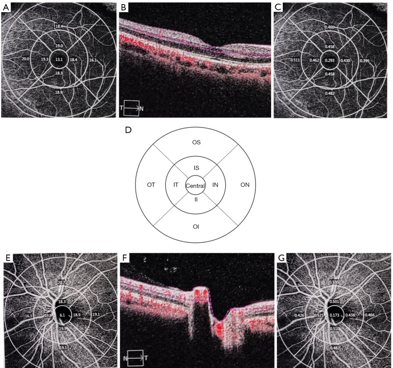
Methods: This is a case-control study. Fifteen patients with IgAN and 15 healthy controls (HCs) who attended The Affiliated Hospital of Southwest Medical University between July 2022 and December 2023 were selected. OCT and OCTA were used to assess macular thickness, retinal nerve fiber layer (RNFL) thickness, the vascular density (VD) and perfusion density (PD) of macular and optic disc, and foveal avascular zone (FAZ). In addition, we analyzed the association between OCT and OCTA parameters and clinical data in patients with IgAN.
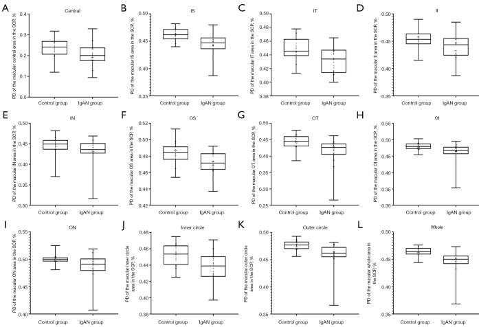
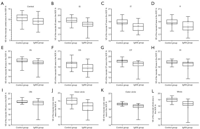
Results: VD and PD were significantly lower in the macular area in the IgAN group than the HCs (VD: 18.270±0.683 vs. 18.943±0.357, P<0.001; PD: 0.447±0.019 vs. 0.464±0.009, P<0.001). VD and PD in the macular area were negatively correlated with urine protein levels (VD: ρ=-0.604, P=0.017; PD: ρ=-0.551, P=0.033) and urine microalbumin levels (VD: ρ=-0.762, P=0.001; PD: ρ=-0.751, P=0.001). The FAZ area was positively correlated with 24-h urine volume (ρ=0.651, P=0.009).
Conclusions: Compared with HCs, the status of macular microcirculation in the IgAN group was significantly decreased, and some microvascular parameters were significantly correlated with the clinical data.

 求助内容:
求助内容: 应助结果提醒方式:


