{"title":"经皮超声造影在乳腺癌前哨淋巴结检测诊断中的价值:与病理特征的比较","authors":"Caixin Huang, Jia Luo, Manying Li, Zhen Shan, Tiantian Zhen, Jiaping Li, Jinyu Liang, Xiaoyan Xie, Yanling Zheng","doi":"10.21037/qims-2024-2631","DOIUrl":null,"url":null,"abstract":"<p><strong>Background: </strong>The diagnostic performance of percutaneous contrast-enhanced ultrasound (P-CEUS) in classifying sentinel lymph nodes (SLNs) in breast cancer varies. This study aimed to evaluate the diagnostic value of P-CEUS in identifying SLNs and to explore the correlation between P-CEUS patterns and pathological characteristics of SLNs.</p><p><strong>Methods: </strong>This retrospective study included consecutive breast cancer patients who underwent preoperative or axillary surgery between June 2019 and March 2021. Each patient underwent conventional ultrasound of the ipsilateral breast and axilla, and SLNs were localized and diagnosed via P-CEUS. Histopathological examination served as the gold standard to assess the diagnostic accuracy of P-CEUS classification based on lymph node structure. The relationship between P-CEUS patterns and pathological characteristics of SLNs was also examined.</p><p><strong>Results: </strong>A total of 238 breast cancer patients (including 1 male) with a mean age of 51.0±11.3 years were included. Five patients with bilateral breast cancer underwent P-CEUS on both sides. The SLN detection rate by P-CEUS was 96.29% (234/243). Pathology results indicated that 64 SLNs (21.8%) were metastatic and 229 SLNs (78.2%) were non-metastatic. The sensitivity, specificity, positive predictive value, negative predictive value, and accuracy of P-CEUS in detecting SLNs were 76.56% (49/64), 97.38% (223/229), 89.09% (49/55), 93.70% (223/238), and 92.83% (272/293), respectively. SLNs with different P-CEUS patterns exhibited varying pathological characteristics. Pathology sections of non-metastatic lymph nodes revealed varying degrees of adipose tissue and fibrous tissue hyperplasia. The different enhancement patterns of metastatic lymph nodes were closely associated with the distribution of metastatic lesions within the lymph nodes.</p><p><strong>Conclusions: </strong>The P-CEUS pattern of SLNs was closely correlated with their pathological characteristics. The P-CEUS classification of SLNs based on lymph node structure could aid in the diagnosis of SLNs in breast cancer.</p>","PeriodicalId":54267,"journal":{"name":"Quantitative Imaging in Medicine and Surgery","volume":"15 9","pages":"8309-8319"},"PeriodicalIF":2.3000,"publicationDate":"2025-09-01","publicationTypes":"Journal Article","fieldsOfStudy":null,"isOpenAccess":false,"openAccessPdf":"https://www.ncbi.nlm.nih.gov/pmc/articles/PMC12397642/pdf/","citationCount":"0","resultStr":"{\"title\":\"The value of percutaneous contrast-enhanced ultrasound in the detection and diagnosis of sentinel lymph nodes in breast cancer: comparison with pathological features.\",\"authors\":\"Caixin Huang, Jia Luo, Manying Li, Zhen Shan, Tiantian Zhen, Jiaping Li, Jinyu Liang, Xiaoyan Xie, Yanling Zheng\",\"doi\":\"10.21037/qims-2024-2631\",\"DOIUrl\":null,\"url\":null,\"abstract\":\"<p><strong>Background: </strong>The diagnostic performance of percutaneous contrast-enhanced ultrasound (P-CEUS) in classifying sentinel lymph nodes (SLNs) in breast cancer varies. This study aimed to evaluate the diagnostic value of P-CEUS in identifying SLNs and to explore the correlation between P-CEUS patterns and pathological characteristics of SLNs.</p><p><strong>Methods: </strong>This retrospective study included consecutive breast cancer patients who underwent preoperative or axillary surgery between June 2019 and March 2021. Each patient underwent conventional ultrasound of the ipsilateral breast and axilla, and SLNs were localized and diagnosed via P-CEUS. Histopathological examination served as the gold standard to assess the diagnostic accuracy of P-CEUS classification based on lymph node structure. The relationship between P-CEUS patterns and pathological characteristics of SLNs was also examined.</p><p><strong>Results: </strong>A total of 238 breast cancer patients (including 1 male) with a mean age of 51.0±11.3 years were included. Five patients with bilateral breast cancer underwent P-CEUS on both sides. The SLN detection rate by P-CEUS was 96.29% (234/243). Pathology results indicated that 64 SLNs (21.8%) were metastatic and 229 SLNs (78.2%) were non-metastatic. The sensitivity, specificity, positive predictive value, negative predictive value, and accuracy of P-CEUS in detecting SLNs were 76.56% (49/64), 97.38% (223/229), 89.09% (49/55), 93.70% (223/238), and 92.83% (272/293), respectively. SLNs with different P-CEUS patterns exhibited varying pathological characteristics. Pathology sections of non-metastatic lymph nodes revealed varying degrees of adipose tissue and fibrous tissue hyperplasia. The different enhancement patterns of metastatic lymph nodes were closely associated with the distribution of metastatic lesions within the lymph nodes.</p><p><strong>Conclusions: </strong>The P-CEUS pattern of SLNs was closely correlated with their pathological characteristics. The P-CEUS classification of SLNs based on lymph node structure could aid in the diagnosis of SLNs in breast cancer.</p>\",\"PeriodicalId\":54267,\"journal\":{\"name\":\"Quantitative Imaging in Medicine and Surgery\",\"volume\":\"15 9\",\"pages\":\"8309-8319\"},\"PeriodicalIF\":2.3000,\"publicationDate\":\"2025-09-01\",\"publicationTypes\":\"Journal Article\",\"fieldsOfStudy\":null,\"isOpenAccess\":false,\"openAccessPdf\":\"https://www.ncbi.nlm.nih.gov/pmc/articles/PMC12397642/pdf/\",\"citationCount\":\"0\",\"resultStr\":null,\"platform\":\"Semanticscholar\",\"paperid\":null,\"PeriodicalName\":\"Quantitative Imaging in Medicine and Surgery\",\"FirstCategoryId\":\"3\",\"ListUrlMain\":\"https://doi.org/10.21037/qims-2024-2631\",\"RegionNum\":2,\"RegionCategory\":\"医学\",\"ArticlePicture\":[],\"TitleCN\":null,\"AbstractTextCN\":null,\"PMCID\":null,\"EPubDate\":\"2025/8/14 0:00:00\",\"PubModel\":\"Epub\",\"JCR\":\"Q2\",\"JCRName\":\"RADIOLOGY, NUCLEAR MEDICINE & MEDICAL IMAGING\",\"Score\":null,\"Total\":0}","platform":"Semanticscholar","paperid":null,"PeriodicalName":"Quantitative Imaging in Medicine and Surgery","FirstCategoryId":"3","ListUrlMain":"https://doi.org/10.21037/qims-2024-2631","RegionNum":2,"RegionCategory":"医学","ArticlePicture":[],"TitleCN":null,"AbstractTextCN":null,"PMCID":null,"EPubDate":"2025/8/14 0:00:00","PubModel":"Epub","JCR":"Q2","JCRName":"RADIOLOGY, NUCLEAR MEDICINE & MEDICAL IMAGING","Score":null,"Total":0}
The value of percutaneous contrast-enhanced ultrasound in the detection and diagnosis of sentinel lymph nodes in breast cancer: comparison with pathological features.
Background: The diagnostic performance of percutaneous contrast-enhanced ultrasound (P-CEUS) in classifying sentinel lymph nodes (SLNs) in breast cancer varies. This study aimed to evaluate the diagnostic value of P-CEUS in identifying SLNs and to explore the correlation between P-CEUS patterns and pathological characteristics of SLNs.
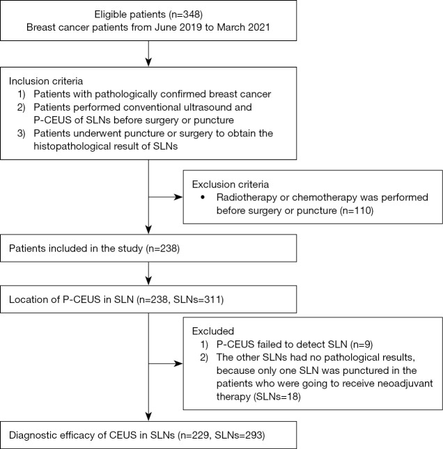
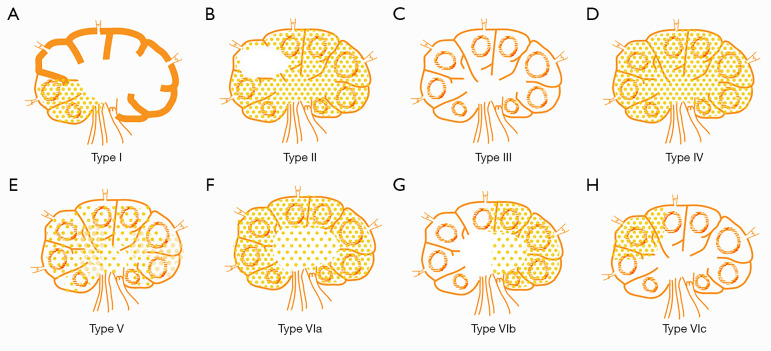
Methods: This retrospective study included consecutive breast cancer patients who underwent preoperative or axillary surgery between June 2019 and March 2021. Each patient underwent conventional ultrasound of the ipsilateral breast and axilla, and SLNs were localized and diagnosed via P-CEUS. Histopathological examination served as the gold standard to assess the diagnostic accuracy of P-CEUS classification based on lymph node structure. The relationship between P-CEUS patterns and pathological characteristics of SLNs was also examined.
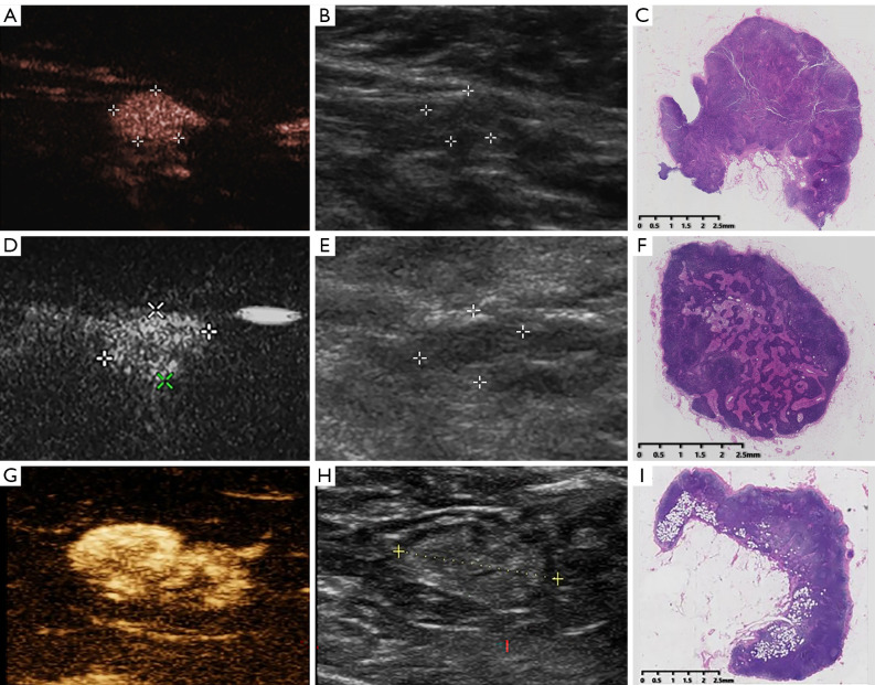
Results: A total of 238 breast cancer patients (including 1 male) with a mean age of 51.0±11.3 years were included. Five patients with bilateral breast cancer underwent P-CEUS on both sides. The SLN detection rate by P-CEUS was 96.29% (234/243). Pathology results indicated that 64 SLNs (21.8%) were metastatic and 229 SLNs (78.2%) were non-metastatic. The sensitivity, specificity, positive predictive value, negative predictive value, and accuracy of P-CEUS in detecting SLNs were 76.56% (49/64), 97.38% (223/229), 89.09% (49/55), 93.70% (223/238), and 92.83% (272/293), respectively. SLNs with different P-CEUS patterns exhibited varying pathological characteristics. Pathology sections of non-metastatic lymph nodes revealed varying degrees of adipose tissue and fibrous tissue hyperplasia. The different enhancement patterns of metastatic lymph nodes were closely associated with the distribution of metastatic lesions within the lymph nodes.
Conclusions: The P-CEUS pattern of SLNs was closely correlated with their pathological characteristics. The P-CEUS classification of SLNs based on lymph node structure could aid in the diagnosis of SLNs in breast cancer.

 求助内容:
求助内容: 应助结果提醒方式:


