Yao Chen, Yuzhen Xi, Fanfan Zhao, Huanhuan Li, Min Zhou, Yue Xu, Shufeng Fan, Miao Liu
{"title":"药物性肝损伤不同类型及严重程度的ct表现。","authors":"Yao Chen, Yuzhen Xi, Fanfan Zhao, Huanhuan Li, Min Zhou, Yue Xu, Shufeng Fan, Miao Liu","doi":"10.21037/qims-24-2110","DOIUrl":null,"url":null,"abstract":"<p><strong>Background: </strong>Drug-induced liver injury (DILI) has become a major cause of acute liver failure, and its incidence has been increasing steadily in recent years. This study aimed to compare the clinical and computed tomography (CT) imaging features of the variable biochemical damage and severity of DILI to establish a radiological model for predicting high-risk DILI based on CT image features.</p><p><strong>Methods: </strong>The eligible patients with DILI (January 2016 to March 2024) who underwent serum laboratory examination and contrast abdominal CT within 3 months of onset were retrospectively analyzed at Affiliated Xihu Hospital of Hangzhou Medical College (Institution I) and The Second Affiliated Hospital of Zhejiang Chinese Medical University (Institution II). The severity-associated CT features were determined via binomial logistic regression analysis, and the efficacy of the different models were compared. The odds ratios (ORs) and corresponding 95% confidence intervals (CIs) provided were not adjusted.</p><p><strong>Results: </strong>The injury types included hepatocellular (n=68, 45.64%), mixed (n=28, 18.79%), and cholestatic (n=53, 35.57%). The proportion of splenomegaly in patients with cholestatic injury (56.60%) was significantly higher than that in those with hepatocellular (35.71%) and mixed injury (22.06%) (P<0.001). Regarding severity, 127 (85.23%) patients had mild-to-moderate injury, and 22 (14.77%) had severe-to-fatal injury or required liver transplantation (LT). Injury severity was independently associated with quantitative liver-spleen contrast (Q-LSC) (OR =0.002; 95% CI: 0.00-0.13), and ascites (OR =70.83; 95% CI: 16.34-306.99). The prediction of the new model employing Q-LSC and ascites for high-risk DILI demonstrated excellent performance [area under the receiver operating characteristic (ROC) curve (AUC) =0.929; sensitivity=0.818; specificity =0.953].</p><p><strong>Conclusions: </strong>Statistical differences are observed in the serum biomarkers of DILI according to varying biochemical damage and degree of severity. Q-LSC and ascites were associated with the severity of DILI, and a combined model incorporating Q-LSC and ascites can effectively predict high-risk DILI.</p>","PeriodicalId":54267,"journal":{"name":"Quantitative Imaging in Medicine and Surgery","volume":"15 9","pages":"8553-8566"},"PeriodicalIF":2.3000,"publicationDate":"2025-09-01","publicationTypes":"Journal Article","fieldsOfStudy":null,"isOpenAccess":false,"openAccessPdf":"https://www.ncbi.nlm.nih.gov/pmc/articles/PMC12397697/pdf/","citationCount":"0","resultStr":"{\"title\":\"Computed tomography manifestations of drug-induced liver injury according to type and severity of injury.\",\"authors\":\"Yao Chen, Yuzhen Xi, Fanfan Zhao, Huanhuan Li, Min Zhou, Yue Xu, Shufeng Fan, Miao Liu\",\"doi\":\"10.21037/qims-24-2110\",\"DOIUrl\":null,\"url\":null,\"abstract\":\"<p><strong>Background: </strong>Drug-induced liver injury (DILI) has become a major cause of acute liver failure, and its incidence has been increasing steadily in recent years. This study aimed to compare the clinical and computed tomography (CT) imaging features of the variable biochemical damage and severity of DILI to establish a radiological model for predicting high-risk DILI based on CT image features.</p><p><strong>Methods: </strong>The eligible patients with DILI (January 2016 to March 2024) who underwent serum laboratory examination and contrast abdominal CT within 3 months of onset were retrospectively analyzed at Affiliated Xihu Hospital of Hangzhou Medical College (Institution I) and The Second Affiliated Hospital of Zhejiang Chinese Medical University (Institution II). The severity-associated CT features were determined via binomial logistic regression analysis, and the efficacy of the different models were compared. The odds ratios (ORs) and corresponding 95% confidence intervals (CIs) provided were not adjusted.</p><p><strong>Results: </strong>The injury types included hepatocellular (n=68, 45.64%), mixed (n=28, 18.79%), and cholestatic (n=53, 35.57%). The proportion of splenomegaly in patients with cholestatic injury (56.60%) was significantly higher than that in those with hepatocellular (35.71%) and mixed injury (22.06%) (P<0.001). Regarding severity, 127 (85.23%) patients had mild-to-moderate injury, and 22 (14.77%) had severe-to-fatal injury or required liver transplantation (LT). Injury severity was independently associated with quantitative liver-spleen contrast (Q-LSC) (OR =0.002; 95% CI: 0.00-0.13), and ascites (OR =70.83; 95% CI: 16.34-306.99). The prediction of the new model employing Q-LSC and ascites for high-risk DILI demonstrated excellent performance [area under the receiver operating characteristic (ROC) curve (AUC) =0.929; sensitivity=0.818; specificity =0.953].</p><p><strong>Conclusions: </strong>Statistical differences are observed in the serum biomarkers of DILI according to varying biochemical damage and degree of severity. Q-LSC and ascites were associated with the severity of DILI, and a combined model incorporating Q-LSC and ascites can effectively predict high-risk DILI.</p>\",\"PeriodicalId\":54267,\"journal\":{\"name\":\"Quantitative Imaging in Medicine and Surgery\",\"volume\":\"15 9\",\"pages\":\"8553-8566\"},\"PeriodicalIF\":2.3000,\"publicationDate\":\"2025-09-01\",\"publicationTypes\":\"Journal Article\",\"fieldsOfStudy\":null,\"isOpenAccess\":false,\"openAccessPdf\":\"https://www.ncbi.nlm.nih.gov/pmc/articles/PMC12397697/pdf/\",\"citationCount\":\"0\",\"resultStr\":null,\"platform\":\"Semanticscholar\",\"paperid\":null,\"PeriodicalName\":\"Quantitative Imaging in Medicine and Surgery\",\"FirstCategoryId\":\"3\",\"ListUrlMain\":\"https://doi.org/10.21037/qims-24-2110\",\"RegionNum\":2,\"RegionCategory\":\"医学\",\"ArticlePicture\":[],\"TitleCN\":null,\"AbstractTextCN\":null,\"PMCID\":null,\"EPubDate\":\"2025/8/11 0:00:00\",\"PubModel\":\"Epub\",\"JCR\":\"Q2\",\"JCRName\":\"RADIOLOGY, NUCLEAR MEDICINE & MEDICAL IMAGING\",\"Score\":null,\"Total\":0}","platform":"Semanticscholar","paperid":null,"PeriodicalName":"Quantitative Imaging in Medicine and Surgery","FirstCategoryId":"3","ListUrlMain":"https://doi.org/10.21037/qims-24-2110","RegionNum":2,"RegionCategory":"医学","ArticlePicture":[],"TitleCN":null,"AbstractTextCN":null,"PMCID":null,"EPubDate":"2025/8/11 0:00:00","PubModel":"Epub","JCR":"Q2","JCRName":"RADIOLOGY, NUCLEAR MEDICINE & MEDICAL IMAGING","Score":null,"Total":0}
Computed tomography manifestations of drug-induced liver injury according to type and severity of injury.
Background: Drug-induced liver injury (DILI) has become a major cause of acute liver failure, and its incidence has been increasing steadily in recent years. This study aimed to compare the clinical and computed tomography (CT) imaging features of the variable biochemical damage and severity of DILI to establish a radiological model for predicting high-risk DILI based on CT image features.
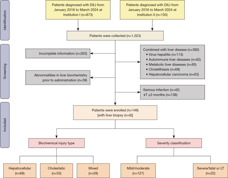
Methods: The eligible patients with DILI (January 2016 to March 2024) who underwent serum laboratory examination and contrast abdominal CT within 3 months of onset were retrospectively analyzed at Affiliated Xihu Hospital of Hangzhou Medical College (Institution I) and The Second Affiliated Hospital of Zhejiang Chinese Medical University (Institution II). The severity-associated CT features were determined via binomial logistic regression analysis, and the efficacy of the different models were compared. The odds ratios (ORs) and corresponding 95% confidence intervals (CIs) provided were not adjusted.
Results: The injury types included hepatocellular (n=68, 45.64%), mixed (n=28, 18.79%), and cholestatic (n=53, 35.57%). The proportion of splenomegaly in patients with cholestatic injury (56.60%) was significantly higher than that in those with hepatocellular (35.71%) and mixed injury (22.06%) (P<0.001). Regarding severity, 127 (85.23%) patients had mild-to-moderate injury, and 22 (14.77%) had severe-to-fatal injury or required liver transplantation (LT). Injury severity was independently associated with quantitative liver-spleen contrast (Q-LSC) (OR =0.002; 95% CI: 0.00-0.13), and ascites (OR =70.83; 95% CI: 16.34-306.99). The prediction of the new model employing Q-LSC and ascites for high-risk DILI demonstrated excellent performance [area under the receiver operating characteristic (ROC) curve (AUC) =0.929; sensitivity=0.818; specificity =0.953].
Conclusions: Statistical differences are observed in the serum biomarkers of DILI according to varying biochemical damage and degree of severity. Q-LSC and ascites were associated with the severity of DILI, and a combined model incorporating Q-LSC and ascites can effectively predict high-risk DILI.


 求助内容:
求助内容: 应助结果提醒方式:


