{"title":"双能计算机断层扫描衍生的电子密度在局灶性肝脏病变鉴别诊断中的初步经验。","authors":"Takashi Ohtani, Kumi Ozaki, Tomokazu Ishida, Kouki Takahashi, Masato Shimada, Satomi Kanai, Tasuku Wakabayashi, Kenji Takata, Toshiki Tateishi, Tetsuya Tsujikawa","doi":"10.21037/qims-2024-2592","DOIUrl":null,"url":null,"abstract":"<p><strong>Background: </strong>The clinical utility of electron density (ED), obtained by non-contrast dual-energy computed tomography, has been demonstrated for the diagnosis of brain tumors and bone lesions; however, the clinical utility of ED in the liver has not been adequately reported. This study aimed to compare ED between hepatocellular carcinomas (HCCs), liver metastases, hepatic hemangiomas, and hepatic cysts and assess the differential diagnostic performance of ED between malignant tumors and benign lesions.</p><p><strong>Methods: </strong>Eighty-nine patients (53 men and 36 women; mean age, 67.2±13.2 years) were included. EDs were measured for 24 HCCs, 19 liver metastases, 28 hepatic hemangiomas, and 18 hepatic cysts. The relative ED (rED), normalized by the ED of the background liver parenchyma, was calculated. The ED and rED of the focal hepatic lesions (FHLs) were statistically analyzed. Receiver operating characteristic curve analysis was used to evaluate the diagnostic performance of ED and rED in differentiating malignant tumors from benign lesions.</p><p><strong>Results: </strong>The ED and rED were higher in malignant tumors than in benign lesions. Significant differences in ED were observed among all lesions (P<0.05) except liver metastases and hepatic hemangiomas. Significant differences in rED were observed among all lesions (P<0.05) except HCCs and liver metastases. The area under the curve of rED for distinguishing between malignant tumors and benign lesions was significantly larger than that of ED (0.88 <i>vs.</i> 0.80, P<0.05).</p><p><strong>Conclusions: </strong>ED aids in the differential diagnosis of FHLs. The use of rED further improves diagnostic performance.</p>","PeriodicalId":54267,"journal":{"name":"Quantitative Imaging in Medicine and Surgery","volume":"15 9","pages":"8219-8229"},"PeriodicalIF":2.3000,"publicationDate":"2025-09-01","publicationTypes":"Journal Article","fieldsOfStudy":null,"isOpenAccess":false,"openAccessPdf":"https://www.ncbi.nlm.nih.gov/pmc/articles/PMC12397626/pdf/","citationCount":"0","resultStr":"{\"title\":\"Dual-energy computed tomography-derived electron density in the differential diagnosis of focal hepatic lesions: initial experience.\",\"authors\":\"Takashi Ohtani, Kumi Ozaki, Tomokazu Ishida, Kouki Takahashi, Masato Shimada, Satomi Kanai, Tasuku Wakabayashi, Kenji Takata, Toshiki Tateishi, Tetsuya Tsujikawa\",\"doi\":\"10.21037/qims-2024-2592\",\"DOIUrl\":null,\"url\":null,\"abstract\":\"<p><strong>Background: </strong>The clinical utility of electron density (ED), obtained by non-contrast dual-energy computed tomography, has been demonstrated for the diagnosis of brain tumors and bone lesions; however, the clinical utility of ED in the liver has not been adequately reported. This study aimed to compare ED between hepatocellular carcinomas (HCCs), liver metastases, hepatic hemangiomas, and hepatic cysts and assess the differential diagnostic performance of ED between malignant tumors and benign lesions.</p><p><strong>Methods: </strong>Eighty-nine patients (53 men and 36 women; mean age, 67.2±13.2 years) were included. EDs were measured for 24 HCCs, 19 liver metastases, 28 hepatic hemangiomas, and 18 hepatic cysts. The relative ED (rED), normalized by the ED of the background liver parenchyma, was calculated. The ED and rED of the focal hepatic lesions (FHLs) were statistically analyzed. Receiver operating characteristic curve analysis was used to evaluate the diagnostic performance of ED and rED in differentiating malignant tumors from benign lesions.</p><p><strong>Results: </strong>The ED and rED were higher in malignant tumors than in benign lesions. Significant differences in ED were observed among all lesions (P<0.05) except liver metastases and hepatic hemangiomas. Significant differences in rED were observed among all lesions (P<0.05) except HCCs and liver metastases. The area under the curve of rED for distinguishing between malignant tumors and benign lesions was significantly larger than that of ED (0.88 <i>vs.</i> 0.80, P<0.05).</p><p><strong>Conclusions: </strong>ED aids in the differential diagnosis of FHLs. The use of rED further improves diagnostic performance.</p>\",\"PeriodicalId\":54267,\"journal\":{\"name\":\"Quantitative Imaging in Medicine and Surgery\",\"volume\":\"15 9\",\"pages\":\"8219-8229\"},\"PeriodicalIF\":2.3000,\"publicationDate\":\"2025-09-01\",\"publicationTypes\":\"Journal Article\",\"fieldsOfStudy\":null,\"isOpenAccess\":false,\"openAccessPdf\":\"https://www.ncbi.nlm.nih.gov/pmc/articles/PMC12397626/pdf/\",\"citationCount\":\"0\",\"resultStr\":null,\"platform\":\"Semanticscholar\",\"paperid\":null,\"PeriodicalName\":\"Quantitative Imaging in Medicine and Surgery\",\"FirstCategoryId\":\"3\",\"ListUrlMain\":\"https://doi.org/10.21037/qims-2024-2592\",\"RegionNum\":2,\"RegionCategory\":\"医学\",\"ArticlePicture\":[],\"TitleCN\":null,\"AbstractTextCN\":null,\"PMCID\":null,\"EPubDate\":\"2025/8/14 0:00:00\",\"PubModel\":\"Epub\",\"JCR\":\"Q2\",\"JCRName\":\"RADIOLOGY, NUCLEAR MEDICINE & MEDICAL IMAGING\",\"Score\":null,\"Total\":0}","platform":"Semanticscholar","paperid":null,"PeriodicalName":"Quantitative Imaging in Medicine and Surgery","FirstCategoryId":"3","ListUrlMain":"https://doi.org/10.21037/qims-2024-2592","RegionNum":2,"RegionCategory":"医学","ArticlePicture":[],"TitleCN":null,"AbstractTextCN":null,"PMCID":null,"EPubDate":"2025/8/14 0:00:00","PubModel":"Epub","JCR":"Q2","JCRName":"RADIOLOGY, NUCLEAR MEDICINE & MEDICAL IMAGING","Score":null,"Total":0}
Dual-energy computed tomography-derived electron density in the differential diagnosis of focal hepatic lesions: initial experience.
Background: The clinical utility of electron density (ED), obtained by non-contrast dual-energy computed tomography, has been demonstrated for the diagnosis of brain tumors and bone lesions; however, the clinical utility of ED in the liver has not been adequately reported. This study aimed to compare ED between hepatocellular carcinomas (HCCs), liver metastases, hepatic hemangiomas, and hepatic cysts and assess the differential diagnostic performance of ED between malignant tumors and benign lesions.
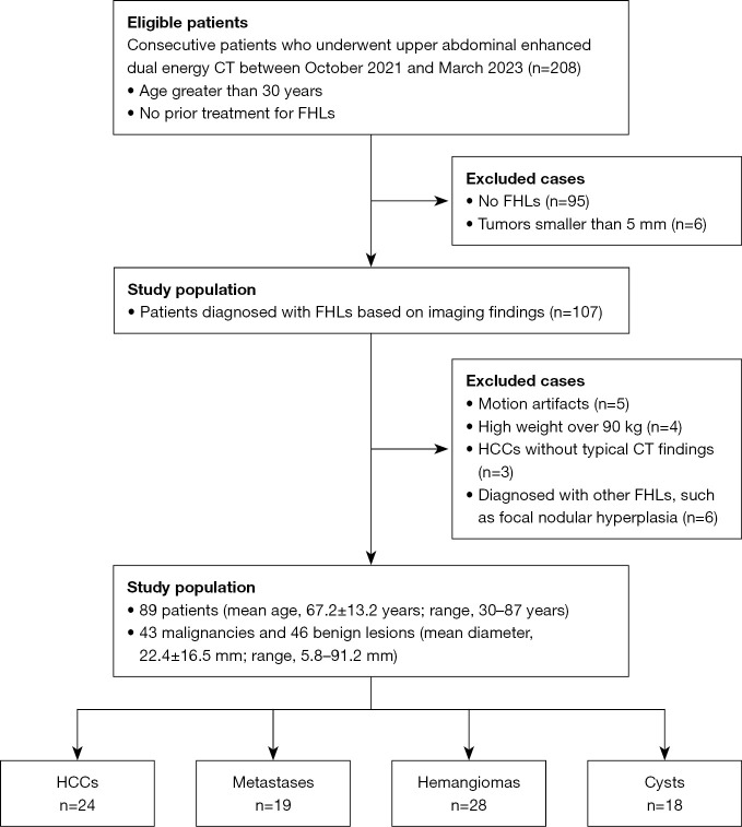
Methods: Eighty-nine patients (53 men and 36 women; mean age, 67.2±13.2 years) were included. EDs were measured for 24 HCCs, 19 liver metastases, 28 hepatic hemangiomas, and 18 hepatic cysts. The relative ED (rED), normalized by the ED of the background liver parenchyma, was calculated. The ED and rED of the focal hepatic lesions (FHLs) were statistically analyzed. Receiver operating characteristic curve analysis was used to evaluate the diagnostic performance of ED and rED in differentiating malignant tumors from benign lesions.
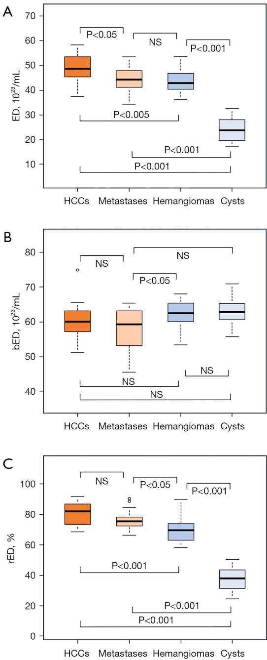
Results: The ED and rED were higher in malignant tumors than in benign lesions. Significant differences in ED were observed among all lesions (P<0.05) except liver metastases and hepatic hemangiomas. Significant differences in rED were observed among all lesions (P<0.05) except HCCs and liver metastases. The area under the curve of rED for distinguishing between malignant tumors and benign lesions was significantly larger than that of ED (0.88 vs. 0.80, P<0.05).
Conclusions: ED aids in the differential diagnosis of FHLs. The use of rED further improves diagnostic performance.

 求助内容:
求助内容: 应助结果提醒方式:


